. 2021 May 6;11(1):9737.
doi: 10.1038/s41598-021-89123-5.

Identification of germ cell-specific Mga variant mRNA that promotes meiosis via impediment of a non-canonical PRC1

Affiliations

Identification of germ cell-specific Mga variant mRNA that promotes meiosis via impediment of a non-canonical PRC1

Yuka Kitamura et al. Sci Rep. .

Abstract

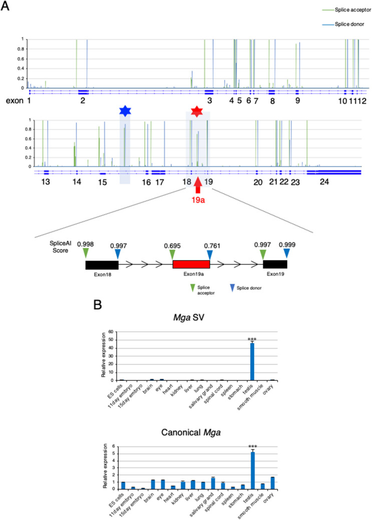
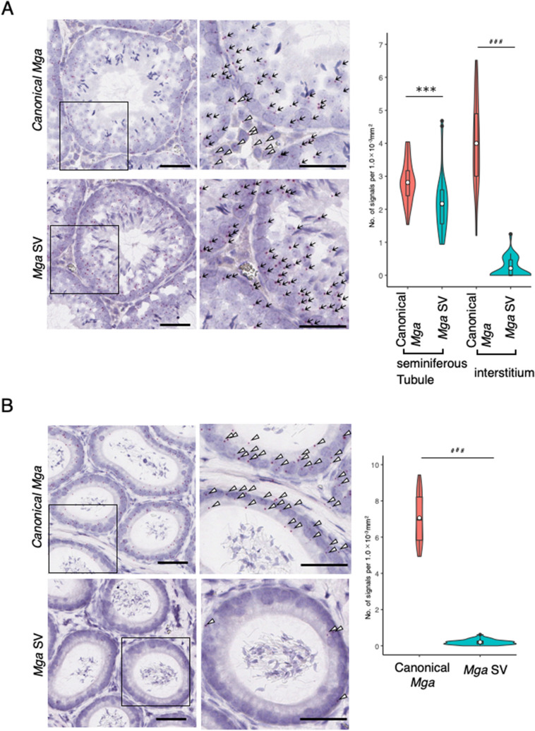
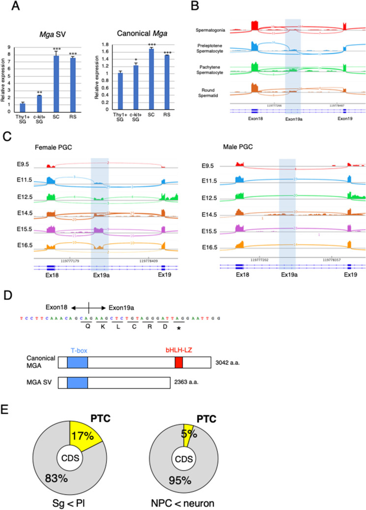
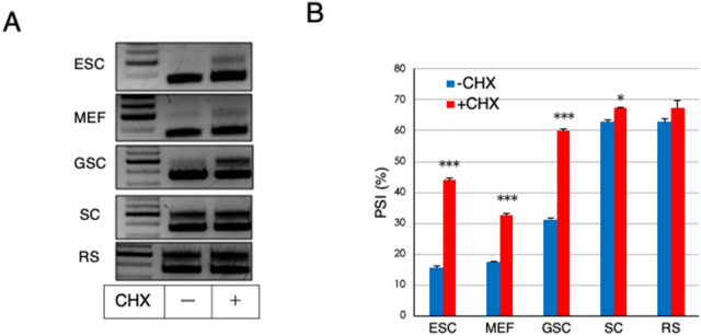
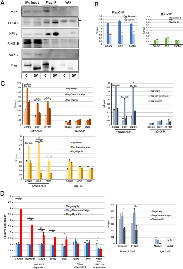
A non-canonical PRC1 (PRC1.6) prevents precocious meiotic onset. Germ cells alleviate its negative effect by reducing their amount of MAX, a component of PRC1.6, as a prerequisite for their bona fide meiosis. Here, we found that germ cells produced Mga variant mRNA bearing a premature termination codon (PTC) during meiosis as an additional mechanism to impede the function of PRC1.6. The variant mRNA encodes an anomalous MGA protein that lacks the bHLHZ domain and thus functions as a dominant negative regulator of PRC1.6. Notwithstanding the presence of PTC, the Mga variant mRNA are rather stably present in spermatocytes and spermatids due to their intrinsic inefficient background of nonsense-mediated mRNA decay. Thus, our data indicate that meiosis is controlled in a multi-layered manner in which both MAX and MGA, which constitute the core of PRC1.6, are at least used as targets to deteriorate the integrity of the complex to ensure progression of meiosis.


Conflict of interest statement

The authors declare no competing interests.

Figures

Figure 1
Figure 1
Identification of testis-specific Mga variant mRNA. (A) Prediction scores as the splice acceptor and donor of Mga pre-mRNA from SpliceAI deep learning. Scores as the splice acceptor and donor are shown as green and blue bars, respectively, whose height is proportional to the score level. In addition to known exons, SpliceAI indicated a set of high scores for the splice acceptor and donor within the regions of the 15th and 18th introns of the Mga gene marked with blue and red asterisks, respectively. The latter region is enlarged to provide actual scores from SpliceAI. (B) qPCR analyses of Mga SV (upper panel) and canonical Mga (lower panel) mRNAs in total RNAs from various tissues. Values obtained from ESCs were arbitrarily set to one for both Mga mRNA species. Data represents the mean ± standard deviation of three independent experiments. The Student’s t-test was conducted to examine statistical significance. ***P < 0.001.
Figure 2
Figure 2
RNA in situ hybridization analyses of canonical and splice variant Mga mRNAs in the testis and epididymis. (A) Detection of canonical mRNA and its splice variant in adult mouse testis by the BaseScope in situ hybridization. The region indicated with an open square is enlarged and shown at the right. Black arrow and open arrowhead indicate transcripts detected within seminiferous tubule (ST) and interstitium (I) portions, respectively. Black bars correspond to 50 μm. Data obtained as the numbers of positive signals per 1.0 mm−3 area in 35 randomly selected areas were used to construct violin plots. For statistical analyses, F-test values were first obtained for the respective data. Then, the Student’s and Welch’s t-tests were conducted when the F-test value was larger and smaller than 0.05, respectively. ***P < 0.001 (Student’s t-test); ###P < 0.001 (Welch’s t-test) (B) Detection of canonical and splice variant Mga mRNAs in the epididymis by the BaseScope in situ hybridization. Detection of canonical Mga mRNA and its splice variant in the adult mouse epididymis and construction of violin plots as described in A. Black bars correspond to 50 μm. Region indicated with an open square is enlarged and shown at the right. Specific signals for canonical (upper panels) and variant (lower panels) Mga mRNAs are indicated with an open arrowhead. Data were analyzed statistically as described in A. ###P < 0.001 (Welch’s t-test).
Figure 3
Figure 3
Mga splice variant is restrictively present around the meiotic stage of germ cells. (A) qPCR analyses of RNAs from undifferentiated (Thy1+) and differentiated (c-Kit+) spermatogonia (SG), spermatocytes (SC), and round spermatids (RS) to determine the levels of Mga SV (left panel) and canonical Mga (right panel) mRNAs. Values obtained from Thy1+ spermatogonia were arbitrarily set to one for both Mga mRNA species. Data were subjected to statistical analyses as described in Fig. 1B. **P < 0.01; ***P < 0.001 (B) Visualization of splicing events around exons 18 and 19 of the Mga gene during spermatogenesis in the testis by Sashimi plots. Plots were constructed using publicly reported data (GSE75826). Abbreviations are described in (A). (C) Sashimi plots showing splicing events around exons 18 and 19 of the Mga gene in male and female PGCs during the embryonic stage. Data for female and male PGCs were obtained from publicly reported data (E-MTAB-4616) and shown in left and right panels, respectively. (D) Sequence of exon 19a containing the in-frame PTC. Acquisition of the exon 19a sequence by alternative splicing was accompanied by insertion of PTC after the addition of the coding sequence of five amino acids, leading to translation into the carboxy-terminally truncated anomalous MGA protein that lacked the bHLHZ domain as depicted in lower portion. (E) Generation of the PTC-containing transcript by alternative splicing was a frequent event during meiotic onset of germ cells. Frequencies of exon inclusion leading to incorporation of a PTC and that leading to addition of the novel amino acid sequence occurred during conversion of spermatogonia to meiotic germ cells (left) and differentiation of neural progenitor cells into neuronal cells (right) are presented as pie charts. Publicly reported RNA sequence data of germ cells (GSE75826) and neuronal cells (GSE96950) were used to calculate the frequencies.
Figure 4
Figure 4
Inefficiency of NMD substantially contributes to accumulation of Mga SV transcripts in meiotic spermatocytes and spermatids. (A) High expression of Mga SV in spermatocytes and round spermatids was not further elevated by suppressing NMD activity. Non-germ (ESC and MEF) and germ (GSC, SC, and RS) cells were treated with or without CHX for 3 h. Semi-quantitative PCR was performed with cDNAs corresponding to their total RNAs. Full-length gels are presented in Supplementary Fig. S7. (B) Quantification of canonical and splice variant Mga mRNAs in non-germ and germ cells treated with or without CHX. cDNAs used in A were subjected to qPCR to accurately compare their amounts. Data are shown as PSI. Data represent the mean ± standard deviation of three independent experiments. The Student’s t-test was conducted to examine statistical significance. *P < 0.05; ***P < 0.001.
Figure 5
Figure 5
Dominant negative effect of carboxy-terminally truncated MGA on PRC1.6. (A) Coimmunoprecipitation analyses of canonical and carboxy-terminally truncated MGAs. Expression vectors for Flag-tagged canonical and carboxy-terminally truncated MGAs were transiently introduced individually into MGA-null HEK293FT cells by transfection. Coimmunoprecipatations were performed with an anti-Flag-tag antibody using nuclear extracts from the transfected cells. Coimmunoprecipitated proteins were used to examine the presence or absence of SUZ12 as well as PRC1.6 components (PCGF6, L3MBTL2, HP1γ, and RING1B). C and SV stand for canonical- and splice variant, respectively. *Indicates signals of the immunoglobulin heavy chain used for immunoprecipitation. Full-length blots are presented in Supplementary Fig. S7. (B) ChIP-qPCR analyses of PRC1.6-target genes in MGA-null HEK293FT cells producing Flag-tagged canonical or carboxy-terminally truncated MGA transiently with the anti-Flag-tag antibody. Control IgG was used as a negative control. Data represent the mean ± standard deviation of three independent experiments. The Student’s t-test was conducted to examine statistical significance. **P < 0.01; ***P < 0.001. (C) ChIP-qPCR analyses of PRC1.6-target genes in MGA-null HEK293FT cells with empty vector and those producing either Flag-tagged canonical or carboxy-terminally truncated MGA transiently with antibodies against MAX, RING1B or PCGF6. Control IgG was used as a negative control. Data represent the mean ± standard deviation of three independent experiments. The Tukey–Kramer test was conducted to examine statistical significance. *P < 0.05; **P < 0.01. (D) Effects of forced expression of Mga SV on meiosis-related genes in mouse ESCs Expression vectors to produce Flag-tagged canonical and carboxy-terminally truncated MGAs and that with no cDNA (empty) were transiently introduced individually into mouse ESCs by transfection. Transfected cells were collected as GFP-positive cells. Then, RNAs prepared from them were used to quantify expression levels of meiosis-related genes (left panel). The same sets of ESCs were also used for ChIP-qPCR analyses of Meiosin and Sycp3 promoter loci with antibody against RING1B (right panel). Control IgG was used as a negative control. N.D. not detected. Data represent the mean ± standard deviation of three independent experiments. Data were subjected to statistical significance examination as in C. *P < 0.05.

Similar articles

Cited by

References

    1. Ginsburg M, Snow MH, McLaren A. Primordial germ cells in the mouse embryo during gastrulation. Development. 1990;110:521–528. - PubMed
    1. Anderson EL, et al. Stra8 and its inducer, retinoic acid, regulate meiotic initiation in both spermatogenesis and oogenesis in mice. Proc. Natl. Acad. Sci. U.S.A. 2008;105:14976–14980. - PMC - PubMed
    1. Ishiguro KI, et al. MEIOSIN directs the switch from mitosis to meiosis in mammalian germ cells. Dev. Cell. 2020;52:429–445. - PubMed
    1. Yokobayashi S, et al. PRC1 coordinates timing of sexual differentiation of female primordial germ cells. Nature. 2013;495:236–240. - PubMed
    1. Maezawa S, et al. Polycomb directs timely activation of germline genes in spermatogenesis. Genes Dev. 2017;31:1693–1703. - PMC - PubMed

Publication types

Substances

LinkOut - more resources