

Circadian disruption by short light exposure and a high energy diet impairs glucose tolerance and increases cardiac fibrosis in Psammomys obesus
- PMID: 33958671
- PMCID: PMC8102519
- DOI: 10.1038/s41598-021-89191-7
Circadian disruption by short light exposure and a high energy diet impairs glucose tolerance and increases cardiac fibrosis in Psammomys obesus
Abstract
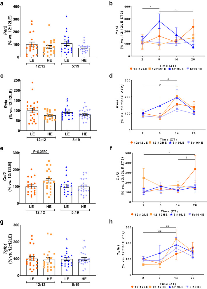
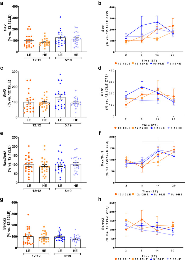
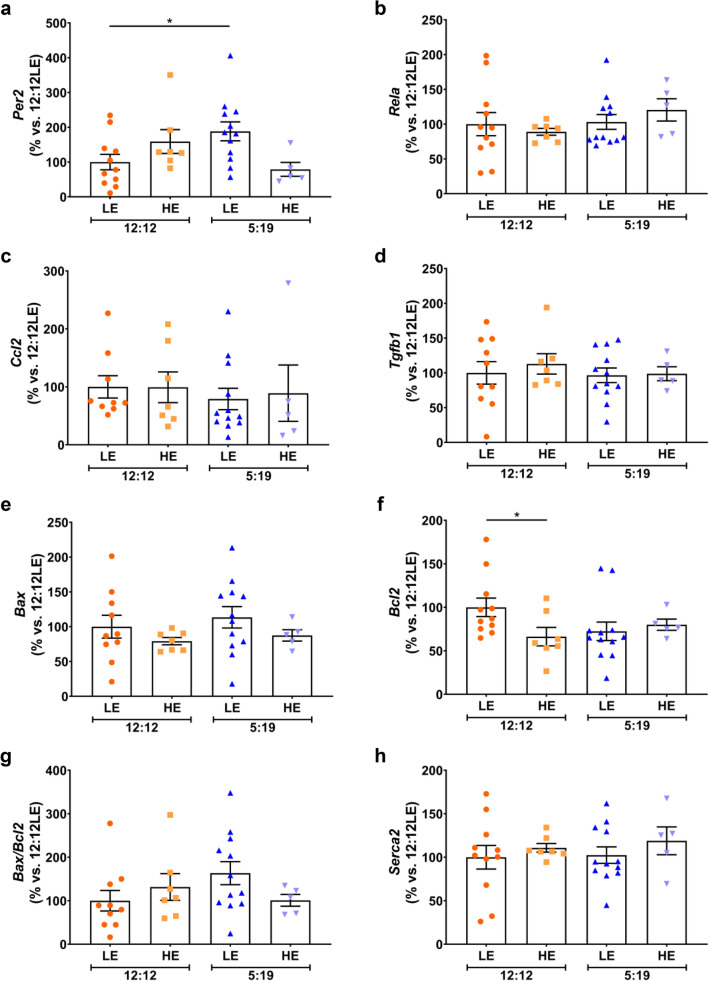
Type 2 diabetes mellitus (T2DM) increases cardiac inflammation which promotes the development of cardiac fibrosis. We sought to determine the impact of circadian disruption on the induction of hyperglycaemia, inflammation and cardiac fibrosis.
Methods: Psammomys obesus (P. obesus) were exposed to neutral (12 h light:12 h dark) or short (5 h light:19 h dark) photoperiods and fed a low energy (LE) or high energy (HE) diet for 8 or 20 weeks. To determine daily rhythmicity, P. obesus were euthanised at 2, 8, 14, and 20 h after 'lights on'.
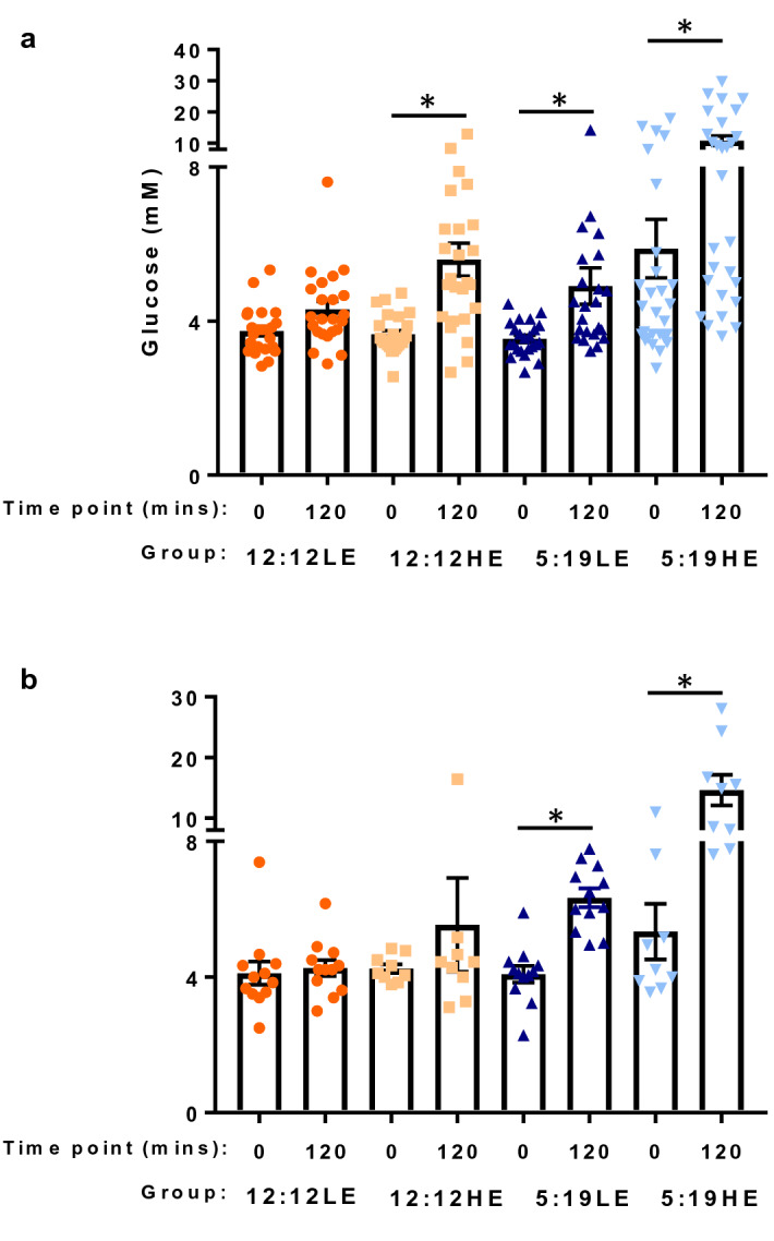
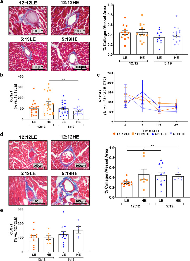
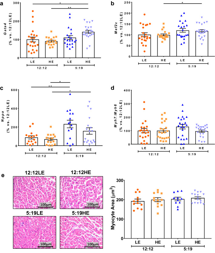
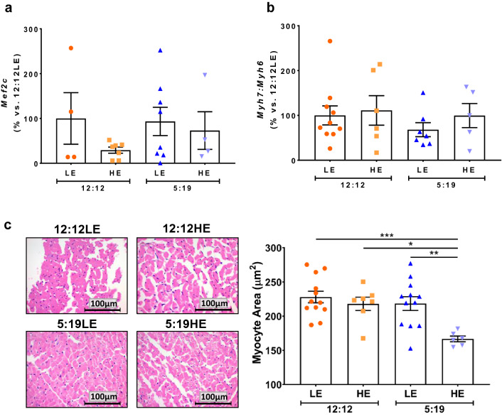
Results: P. obesus exposed to a short photoperiod for 8 and 20 weeks had impaired glucose tolerance following oral glucose tolerance testing, compared to a neutral photoperiod exposure. This occurred with both LE and HE diets but was more pronounced with the HE diet. Short photoperiod exposure also increased myocardial perivascular fibrosis after 20 weeks on LE (51%, P < 0.05) and HE (44%, P < 0.05) diets, when compared to groups with neutral photoperiod exposure. Short photoperiod exposure caused elevations in mRNA levels of hypertrophy gene Nppa (atrial natriuretic peptide) and hypertrophy transcription factors Gata4 and Mef2c in myocardial tissue after 8 weeks.
Conclusion: Exposure to a short photoperiod causes impaired glucose tolerance in P. obesus that is exacerbated with HE diet and is accompanied by an induction in myocardial perivascular fibrosis.
Conflict of interest statement
The authors declare no competing interests.
Figures
Similar articles
-
Female Psammomys obesus Are Protected from Circadian Disruption-Induced Glucose Intolerance, Cardiac Fibrosis and Adipocyte Dysfunction.Int J Mol Sci. 2024 Jul 1;25(13):7265. doi: 10.3390/ijms25137265. Int J Mol Sci. 2024. PMID: 39000372 Free PMC article.
-
High-Energy Diet and Shorter Light Exposure Drives Markers of Adipocyte Dysfunction in Visceral and Subcutaneous Adipose Depots of Psammomys obesus.Int J Mol Sci. 2019 Dec 13;20(24):6291. doi: 10.3390/ijms20246291. Int J Mol Sci. 2019. PMID: 31847097 Free PMC article.
-
Exercise Reduces Glucose Intolerance, Cardiac Inflammation and Adipose Tissue Dysfunction in Psammomys obesus Exposed to Short Photoperiod and High Energy Diet.Int J Mol Sci. 2024 Jul 15;25(14):7756. doi: 10.3390/ijms25147756. Int J Mol Sci. 2024. PMID: 39062999 Free PMC article.
-
Treatment of diabetes with vanadium salts: general overview and amelioration of nutritionally induced diabetes in the Psammomys obesus gerbil.Diabetes Metab Res Rev. 2001 Jan-Feb;17(1):55-66. doi: 10.1002/1520-7560(2000)9999:9999<::aid-dmrr165>3.0.co;2-j. Diabetes Metab Res Rev. 2001. PMID: 11241892 Review.
-
Nutritionally induced diabetes in desert rodents as models of type 2 diabetes: Acomys cahirinus (spiny mice) and Psammomys obesus (desert gerbil).ILAR J. 2006;47(3):212-24. doi: 10.1093/ilar.47.3.212. ILAR J. 2006. PMID: 16804196 Review.
Cited by
-
Circadian rhythms-related disorders in diurnal fat sand rats under modern lifestyle conditions: A review.Front Physiol. 2022 Sep 7;13:963449. doi: 10.3389/fphys.2022.963449. eCollection 2022. Front Physiol. 2022. PMID: 36160856 Free PMC article. Review.
-
A Growing Link between Circadian Rhythms, Type 2 Diabetes Mellitus and Alzheimer's Disease.Int J Mol Sci. 2022 Jan 3;23(1):504. doi: 10.3390/ijms23010504. Int J Mol Sci. 2022. PMID: 35008933 Free PMC article. Review.
-
Effects of photoperiod and food on glucose intolerance and subsequent ocular pathology in the fat sand rat.Sci Rep. 2024 Jan 3;14(1):403. doi: 10.1038/s41598-023-44584-8. Sci Rep. 2024. PMID: 38172147 Free PMC article.
-
Attention to Innate Circadian Rhythm and the Impact of Its Disruption on Diabetes.Diabetes Metab J. 2024 Jan;48(1):37-52. doi: 10.4093/dmj.2023.0193. Epub 2024 Jan 3. Diabetes Metab J. 2024. PMID: 38173377 Free PMC article. Review.
-
Role of circadian rhythms in heart failure: insights from myocardial energy metabolism.J Transl Med. 2025 Jul 10;23(1):770. doi: 10.1186/s12967-025-06828-1. J Transl Med. 2025. PMID: 40640835 Free PMC article. Review.
References
-
- Dunlay SM, et al. Type 2 diabetes mellitus and heart failure: A scientific statement from the American Heart Association and the Heart Failure Society of America: This statement does not represent an update of the 2017 ACC/AHA/HFSA heart failure guideline update. Circulation. 2019;140(7):e294–e324. doi: 10.1161/CIR.0000000000000691. - DOI - PubMed
Publication types
MeSH terms
Substances
LinkOut - more resources
Full Text Sources
Other Literature Sources
Medical
