Newer insights into the clinical profile of posterior lenticonus in children and its surgical, visual, refractive outcomes
- PMID: 33958736
- PMCID: PMC9046400
- DOI: 10.1038/s41433-021-01564-4
Newer insights into the clinical profile of posterior lenticonus in children and its surgical, visual, refractive outcomes
Abstract
Purpose: To analyze the clinical profile of patients with posterior lenticonus and their surgical, visual, and refractive outcomes.
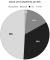
Results: Retrospective interventional case series of 84 eyes of 63 patients with posterior lenticonus. The incidence of posterior lenticonus was 3.98% during a study period of 5 years. One-third of cases had bilateral posterior lenticonus. The mean age was 4.78 ± 4.28 years (unilateral cases were significantly older than bilateral, P = 0.0001). Males were 54%. Mean axial length and keratometry were 21.49 mm and 44.88 D, respectively. Eyes with the bilateral disease were significantly shorter (axial length, P = 0.0012) and smaller (horizontal corneal diameter, P < 0.0001) compared to those with unilateral disease. While 88% were pseudophakic; 12% were aphakic. The posterior capsular defect was noted intraoperatively in 44%. Sixty-eight percent of eyes had a pre-operative diagnosis of posterior lenticonus, 32% were diagnosed intraoperatively. The mean follow-up period was 1.3 years. Best-corrected visual acuity (BCVA) at 6 months was fair to poor in two-third of patients (median 20/100). The mean ± SD visual acuity (LogMAR) and spherical equivalence for unilateral and bilateral cases were 0.70 ± 0.27, 0.67 ± 0.26D (p = 0.57) and 2.04 ± 2.74, 5.15 ± 3.73D (p = 0.0001), respectively. Visual outcomes were better in children who are aged 2 years or more (P = 0.0056). Eight percent needed a second surgery.
Conclusion: We report a higher prevalence of bilateral posterior lenticonus in this cohort. The clinical profile of bilateral disease differs from unilateral disease. The diagnosis is not always clinical. In the bag, intra-ocular lens (IOL) implantation is possible in the majority. The visual outcomes remain fair to poor, possibly due to late presentation and the presence of dense refractory amblyopia.
Synopsis: The manuscript consists of the largest series of posterior lenticonus to date. It provides the prevalence of posterior lenticonus along with characteristics difference between unilateral and bilateral cases of posterior lenticonus. Newer insights in terms of diagnostics, pre-operative pick-up rate, how to improve, visual and refractive outcomes of unilateral and bilateral cases are described.
© 2021. The Author(s), under exclusive licence to The Royal College of Ophthalmologists.
Conflict of interest statement
The authors declare no competing interests.
Figures


References
-
- Mann I. Developmental abnormalities of the eye. Philadelphia: JB Lippincott. 1957:307.
MeSH terms
LinkOut - more resources
Full Text Sources
Other Literature Sources
Medical

