. 2021 Jun;6(6):806-817.
doi: 10.1038/s41564-021-00899-8. Epub 2021 May 6.

A natural symbiotic bacterium drives mosquito refractoriness to Plasmodium infection via secretion of an antimalarial lipase

Affiliations

A natural symbiotic bacterium drives mosquito refractoriness to Plasmodium infection via secretion of an antimalarial lipase

Han Gao et al. Nat Microbiol. 2021 Jun.

Abstract

The stalling global progress in the fight against malaria prompts the urgent need to develop new intervention strategies. Whilst engineered symbiotic bacteria have been shown to confer mosquito resistance to parasite infection, a major challenge for field implementation is to address regulatory concerns. Here, we report the identification of a Plasmodium-blocking symbiotic bacterium, Serratia ureilytica Su_YN1, isolated from the midgut of wild Anopheles sinensis in China that inhibits malaria parasites via secretion of an antimalarial lipase. Analysis of Plasmodium vivax epidemic data indicates that local malaria cases in Tengchong (Yunnan province, China) are significantly lower than imported cases and importantly, that the local vector A. sinensis is more resistant to infection by P. vivax than A. sinensis from other regions. Analysis of the gut symbiotic bacteria of mosquitoes from Yunnan province led to the identification of S. ureilytica Su_YN1. This bacterium renders mosquitoes resistant to infection by the human parasite Plasmodium falciparum or the rodent parasite Plasmodium berghei via secretion of a lipase that selectively kills parasites at various stages. Importantly, Su_YN1 rapidly disseminates through mosquito populations by vertical and horizontal transmission, providing a potential tool for blocking malaria transmission in the field.


Conflict of interest statement

Competing interests

The authors declare no competing interests.

Figures

Extended Data Fig. 1 |
Extended Data Fig. 1 |. Persistence of non-symbiotic bacteria in mosquito midguts.
GFP-tagged E. coli and S. aureus were administered to newly emerged female mosquitoes via sugar feeding. Bacteria midgut colonization was determined by plating serially diluted midgut homogenates on LB agar plates containing 100 μg/ml kanamycin. a,b, E. coli and S. aureus bacteria numbers in mosquitoes maintained with sugar. c,d, E. coli and S. aureus bacteria numbers in mosquitoes post a blood meal. Data points are mean ± s.d. The dots represent biologically independent replicates (n = 2). Each replicate contains 10 mosquito midguts.
Extended Data Fig. 2 |
Extended Data Fig. 2 |. Serratia strains stably colonize the mosquito midgut and do not impact mosquito longevity.
a, b, Effect of Serratia strains on An. sinensis (a) and An. stephensi (b) survival post blood meal. Bacteria were administered to two-day-old female mosquitoes via sugar meal and then fed blood. Survival was monitored daily. Data points are mean ± s.d. The dots represent biologically independent replicates (n = 2). Each replicate contains 10 mosquito midguts. c,d, Serratia bacteria numbers in the midgut of female An. sinensis (c) and An. stephensi (d) post blood meal. Data points are mean ± s.d. The dots represent biologically independent replicates (n = 2). Each replicate contains 10 mosquito midguts. e, Visualization of GFP-tagged Su_YN1 and Sm_YN3 bacteria in the midgut of An. stephensi at 24 h after a blood meal. Bright-field images (left) are paired with the corresponding fluorescent images (right). The experiments were repeated twice with similar results.
Extended Data Fig. 3 |
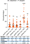
Extended Data Fig. 3 |. Effect of bacteria on P. berghei oocyst formation in An. stephensi.
a, P. berghei oocyst load in An. stephensi mosquitoes carrying Asaia, Acinetobacter or Pantoea bacteria from YN wild caught mosquitoes. b, P. berghei oocyst load in An. stephensi mosquitoes fed with different Serratia strains from different wild-caught mosquito populations. Circles represent the number of oocysts in individual midguts, and horizontal lines indicate the median number of oocysts per midgut. The sample size (n Number) of each group is listed in the table of the lower panel. The statistical significance of the oocyst intensity between the bacteria-fed mosquitoes and PBS-fed mosquitoes (Ctrl) was analysed using the two-tailed Mann-Whitney test. ****P < 0.0001, P > 0.05, not significant (ns). The exact P values in (a) were as follows: Asaia, 0.4508; Acinetobacter, 0.8728; Pantoea, 0.4265. The exact P values in (b) were as follows: Su_YN1, < 0.0001; Sm_YN3, < 0.0001; Sf_JS1, 0.0271; Sf_JS2, 0.0135; Su_JS3, 0.0117; Sm_LN1, 0.0220.
Extended Data Fig. 4 |
Extended Data Fig. 4 |. The effect of Serratia Su_YN1 and Sm_YN3 on An. stephensi blood feeding, fecundity and oviposition rate.
Serratia Su_YN1 and Sm_YN3 do not impact An. stephensi mosquito blood feeding behaviour (n = 100 mosquitoes each group) (a), egg production (Ctrl, n = 46, Su_YN1, n = 39, Sm_YN3, n = 44) (b) or oviposition rate (n = 100 mosquitoes each group) c, Two-day-old An. stephensi mosquitoes were fed on a sugar meal containing bacteria or 5% sugar alone (Ctrl). Three days later, female mosquitoes were fed on a mouse and three days later eggs were collected from individual females. The experiments were repeated three times with similar results. No significant differences were detected among the groups (one-way ANOVA or two-tailed Mann-Whitney test).
Extended Data Fig. 5 |
Extended Data Fig. 5 |. Effect of Rel1 and Rel2 silencing on Su_YN1- or Sm_YN3-mediated anti-Plasmodium activity.
Rel1 and Rel2 were silenced in An. stephensi by systemic injection of double-stranded RNA dsRel1, dsRel2 or dsGFP. The injected mosquitoes were fed on a sugar meal containing Su_YN1 or Sm_YN3. Three days later, the mosquitoes were allowed to feed on the same P. berghei infected mouse. The injected double-stranded RNA (ds) and presence of bacteria are indicated below each column. Each dot represents the oocyst number of an individual midgut, and the horizontal lines indicate the median number of oocysts per midgut. Data are from n = 25 to 28 mosquitoes per group. The sample size (n Number) of each group is listed in the table of the lower panel. The statistical significance of oocyst intensity differences between the dsRel1- or dsRel2-injected and dsGFP-injected mosquitoes carrying the same bacteria (Su_YN1 or Sm_YN3) was analysed using the two-tailed Mann-Whitney test. ****P < 0.0001, P > 0.05, not significant (ns). The exact values were as follows: dsRel1+Su_YN1, 0.8519; dsRel2+Su_YN1, 0.5605; dsRel1+Sm_YN3, < 0.0001; sRel2+Sm_YN3, 0.4851.
Extended Data Fig. 6 |
Extended Data Fig. 6 |. Su_YN1 culture supernatant inhibited P. berghei ookinete formation.
a, Inhibition by Serratia culture supernatants of P. yoelii ookinete formation in vitro. Ookinete formation was quantified by luminescence measurements using the Py.17XNL reporter strain. RLU, relative light units. The experiments were repeated three times with similar results. Data points are mean ± s.d. The dots represent biologically independent replicates (n = 2). b, Effect of Su_YN1 culture supernatant on P. berghei ookinete formation in vitro assay using Giemsa staining. Transformation rate was quantified by comparison with the PBS control. The experiments were repeated three times with similar results. Data points are mean ± s.d. The dots represent biologically independent replicates (n = 3). Statistical significance of the ookinete transformation rate was compared with the PBS control using two-tailed Student’s t-test, ****P < 0.0001. The exact P value was, < 0.0001.
Extended Data Fig. 7 |
Extended Data Fig. 7 |. Haemolytic activity assay of Su_YN1 and Sm_YN3.
a, Bacteria culture supernatant was added (10% V/V) and the mixture incubated with erythrocytes for 12 h. b, The supernatants were collected and the absorbance at 540 nm was measured to evaluate haemoglobin release. Saponin was used as a haemolytic positive control. Data points are mean ± s.d. The dots represent biologically independent replicates (n = 3). Statistical significance of haemolytic activity was compared with the PBS control using two-tailed Student’s t-test. P > 0.05, not significant (ns). The exact P values were as follows: Su_YN1, 0.1672; Sm_YN3, 0.3754.
Extended Data Fig. 8 |
Extended Data Fig. 8 |. Antimalarial activity of different Su_YN1 culture supernatant fractions.
a, Antimalarial activity of organic solvent extracts. Su_YN1 culture supernatant was extracted with solvents of various polarities. The extracted fractions were dried and dissolved in DMSO and the remaining aqueous phase was vacuum treated to remove residual organic reagents. All fractions were tested for antimalarial activity. b, Antimalarial activity assay of Su_YN1 culture supernatant separated using a 3 kDa cut-off centrifugal filter. The retentate and the filtrate were tested. c, Trypsin digestion of Su_YN1 culture supernatant abolishes antimalarial activity. Coomassie Brilliant Blue staining in the lower panel shows the protein patterns before and after treatment. Data points in (b) and (c) are mean ± s.d. The dots represent biologically independent replicates (n = 3). Statistical significance of the ookinete inhibition rate was compared with the PBS control using two-tailed Student’s t-test, ****P < 0.0001, P > 0.05, not significant (ns). The exact P values in (b) were: Trypsin, < 0.0001; Trypsin-inactivated, 0.0552. The exact P values in (c) were: Retentate (compared with PBS), < 0.0001; Filtrate (compared with Retentate), < 0.0001.
Extended Data Fig. 9 |
Extended Data Fig. 9 |. AmLip gene expression in different Serratia strains.
Detection of AmLip transcript abundance in different Serratia strains by qRT-PCR using Serratia 16 s rRNA as an internal reference. Data points are mean ± s.d. The dots represent biologically independent replicates (n = 3). The experiments were repeated twice with similar results.
Extended Data Fig. 10 |
Extended Data Fig. 10 |. Synthesis and purification of Serratia AmLip protein.
a, Western blot detection using HA antibody, of 3HA-tagged AmLip protein in bacterial extracts of Su_YN1 wild type, knock-out mutant AmLip-KO and a mutant AmLip-KO complemented with the AmLip:HA gene. The experiments were repeated twice with similar results. b, Knockout of AmLip was verified by Western blot assay using an AmLip mouse antiserum. The experiments were repeated twice with similar results. c, Expression and purification of AmLip protein expressed E. coli BL21 (DE3). Coomassie blue staining showing the AmLip protein before and after purification on a nickel column. The experiments were repeated twice with similar results. d, Lipase activity test of the purified AmLip protein using the egg yolk lipoprotein plate degradation assay. e, Lipase activity assay of the purified AmLip protein using the trioleoylglycerol- rhodamine B plate degradation assay.
Fig. 1 |
Fig. 1 |. Annual P. vivax cases and A. sinensis susceptibility to P. vivax infection.
a,b, Local and imported cases of P. vivax in YN (Tengchong; a) and JS (Wuxi; b) from 2000 to 2016. Statistical significance of the differences between the occurrence of local and imported cases were determined using two-tailed paired Student’s t-tests. a, P < 0.0001; correlation coefficient of pairing (r) = 0.7314. b, P = 0.0852; r = 0.7192. c, Infection prevalence of wild-caught A. sinensis from YN and JS when fed P. vivax-infected blood. d, Susceptibility of wild-caught A. sinensis mosquitoes from YN (n = 31) and JS (n = 55) to P. vivax infection. P. vivax-infected blood from six patients was used to feed the wild-caught A. sinensis mosquitoes. The circles represent the number of oocysts in individual midguts and the horizontal lines indicate the mean. Statistical significance between the two groups was determined using a two-tailed Mann–Whitney test; P = 0.0114; *P < 0.05.
Fig. 2 |
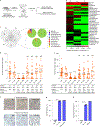
Fig. 2 |. Effect of gut symbiotic Serratia bacteria of wild mosquitoes on Plasmodium development.
a, Workflow of the isolation and identification of mosquito-gut symbiotic bacteria using the gut symbiotic bacteria enrichment method. b, Heat map showing the relative abundance of the top 30 most abundant bacterial genera in A. sinensis mosquito populations from different geographical locations (YN, JS and LN; Supplementary Fig. 1). c, Composition and relative abundance of the genera shared between the three A. sinensis geographical populations. d,e, Effect of different Serratia strains on P. falciparum development in the midgut of A. stephensi (d) and A. gambiae (e) mosquitoes. Female mosquitoes were fed on 5% (wt/vol) sugar solution supplemented with PBS (Ctrl), E. coli or different Serratia strains. Su, S. ureilytica; Sm, S. marcescens; Sf, S. fonticola. The circles represent number of oocysts in individual midguts and the horizontal lines indicate the median. The sample size (number) of each group is listed in the tables (bottom). The experiments were repeated three times with similar results. Statistical significance of the differences in oocyst intensity between the Serratia-fed and E. coli-fed mosquitoes was determined using a two-tailed Mann–Whitney test. f, Images of Giemsa-stained parasites inhibited by the Su_YN1 culture supernatant (related to g,h). Scale bars, 5 μm. The experiments were repeated twice with similar results. g, Inhibition of P. falciparum growth (asexual) by the Su_YN1 culture supernatant. Chloroquine (5 μM) was used as a positive control. h, Inhibition of P. falciparum gametocyte formation by Su_YN1 bacteria in a trans-well assay. The Plasmodium PI4K inhibitor KDU691 (20 μM) was used as a positive control. g,h, The experiments were repeated twice with similar results. Data are the mean ± s.d. The dots represent biologically independent replicates (n = 3). Statistical significance of the inhibition rate compared with the PBS control was determined using a two-tailed Student’s t-test. ****P < 0.0001; NS, not significant (P > 0.05).
Fig. 3 |
Fig. 3 |. Identification of the secreted antimalarial lipase AmLip in S. ureilytica Su_YN1.
a, Comparative analysis of the proteins secreted by the antimalarial S. ureilytica strain Su_YN1 and non-antimalarial S. ureilytica strain Su_JS3. The proteins with higher abundance that were differentially expressed by Su_YN1 and Su_JS3 are displayed; AmLip is highlighted in red font. b, Lipase AmLip is responsible for the antimalarial activity of Su_YN1. The genes coding for the proteins in a were knocked out in Su_YN1 and the culture supernatants of the mutants were used in in vitro ookinete inhibition assays. Two clones of each mutant were tested. Data are the mean ± s.d. The dots represent biologically independent replicates (n = 3). Statistical significance of the test inhibition rate compared with the Su_YN1 wild-type group was determined using a two-tailed Student’s t-test. c, Measurement of lipase enzymatic activity using the lipoprotein plate degradation assay (bottom). Culture supernatants (10 μl) of the indicated bacterial Su_YN1 strains were spotted on an egg yolk plate and incubated for 20 h at 30 °C. Lipase activity was visualized by the lytic spot. The S207A mutation abolished lipase activity. The lipase protein structure is shown with the amino-acid-residue positions indicated (top). d, In vitro Plasmodium yoelii ookinete inhibition assay of the culture supernatants of the indicated Su_YN1 strains. Data are the mean ± s.d. The dots represent biologically independent replicates (n = 3). The experiments were repeated three times with similar results. Statistical significance of the test inhibition rate compared with the wild-type group was determined using a two-tailed Student’s t-test; the P values were as follows: <0.0001 (AmLip-KO, AmLipS207A-comp cl.1 and AmLipS207A-comp cl.2), 0.2224 (AmLip-comp cl.1) and 0.0759 (AmLip-comp cl.2). e, P. berghei ANKA oocyst load in A. stephensi mosquitoes fed with the indicated Su_YN1 strains. The circles represent the number of oocysts in individual midguts and horizontal lines indicate the median. The sample size (number) of each group is listed in the table (bottom). The experiments were repeated twice with similar results. Statistical significance of the difference in oocyst intensity compared with the control group was determined using a two-tailed Mann–Whitney test. ce, AmLip-comp, AmLip complemented strain; AmLipS207A-comp, AmLipS207A point-mutant complemented strain. f, Diagram showing a premature termination codon (indicated in red) in the AmLip coding region of Su_JS3, leading to early termination of translation. g, Western blot detection of AmLip protein in the Su_YN1 and Su_JS3 culture supernatants using anti-AmLip antiserum. The experiments were repeated twice with similar results. ****P < 0.0001; NS, not significant (P > 0.05).
Fig. 4 |
Fig. 4 |. AmLip disrupts and kills parasites.
a, Indirect immunofluorescence detection of AmLip in P. falciparum 3D7 asexual parasites. The 3D7–GFP parasites were incubated with the culture supernatant of 3HA-tagged AmLip (Extended Data Fig. 10a). Binding of AmLip protein to the parasite was detected using HA antibody. The experiments were repeated twice with similar results. BF–Hst, Bright-field images of Hoechst 33342 staining. b, Indirect immunofluorescence detection of AmLip in P. berghei ANKA ookinetes. GFP-labelled ookinetes were incubated with the culture supernatant from wild-type Su_YN1 (bottom) and the AmLip-KO mutant (top). Binding of AmLip protein to the ookinete was detected using AmLip mouse antiserum. The experiments were repeated twice with similar results. c, P. falciparum ookinete load in A. stephensi carrying wild-type Su_YN1 or its AmLip-KO mutant strain. The mosquitoes (n = 10 mosquitoes per group) were fed mature P. falciparum gametocytes, their midguts were dissected 20 h post feeding and the ookinetes in the bolus were counted. The circles represent the number of ookinetes in individual midguts and horizontal lines indicate the median. This experiment was repeated twice with similar results. Statistical significance was analysed using a two-tailed Mann–Whitney test. d, AmLip disrupts the growth and gametocytaemia of P. falciparum parasites. Purified AmLip and AmLipS207A protein were added to P. falciparum 3D7 asexual (top) and gametocyte (bottom) cultures at a final concentration of 5 μg ml−1 and incubated for 24 h. The parasites were observed using Giemsa staining (left) and the per cent inhibition was calculated relative to the PBS controls (right). The experiments were repeated twice with similar results. Data are the mean ± s.d. The dots represent biologically independent replicates (n = 3). Statistical significance of the test inhibition rate compared with the PBS control was determined using a two-tailed Student’s t-test. Top: P = 0.0016 (AmLip) and 0.8817 (AmLipS207A); bottom: P = 0.0001 (AmLip) and 0.6151 (AmLipS207A). e, AmLip antiserum neutralizes the antimalarial activity of the Su_YN1 culture supernatant. Various amounts of AmLip antiserum were added to an ookinete culture. Pre-immune serum was used as a negative control. Data are the mean ± s.d. The dots represent biologically independent replicates (n = 3). Scale bars, 5 μm. ****P < 0.0001; ***P < 0.001; **P < 0.01; NS, not significant (P > 0.05).
Fig. 5 |
Fig. 5 |. S. ureilytica Su_YN1 bacteria spread efficiently through mosquito populations.
a, Su_YN1 is transmitted vertically via laid eggs (n = 21; left), trans-stadially from larvae to adult mosquitoes (n = 20; middle) and venereally from male to female mosquitoes (n = 13; right). The circles represent the number of colony-forming units (c.f.u.) in individual eggs or midguts and horizontal lines indicate the median number of Su_YN1 per egg or midgut. The experiment was repeated three times with similar results. b, Bacterial load in larva (c.f.u. per midgut; n = 14) and breeding water (c.f.u. ml−1) (left) as well as in adult midguts (right; female (n = 20) and male (n = 14)). c, Su_YN1 was transmitted for three consecutive generations. Ten male mosquitoes fed with fluorescent S. ureilytica Su_YN1 bacteria were introduced into a cage with 190 non-bacteria-fed males and 200 virgin females (Supplementary Fig. 7), allowed to mate and the eggs were collected and reared to adults to yield generation F1. Eggs from these adults were reared without further addition of Su_YN1 bacteria to yield F2. F3 was generated similarly. The number of Su_YN1 bacteria in adult female mosquitoes of F1 (n = 10), F2 (n = 13) and F3 (n = 20) were determined. Data are presented as the mean ± s.d. Statistical significance was determined using a one-way ANOVA (without matching or pairing) analysis of variance; P = 0.2887; NS, not significant (P > 0.05). d, Diagram illustrating how Su_YN1 originating from males can spread efficiently through a subsequent mosquito population and be maintained through a complete mosquito life cycle.

Similar articles

Cited by

References

    1. World Malaria Report (World Health Organization, 2019).
    1. Global Vector Control Response 2017–2030 (World Health Organization, 2017).
    1. Dondorp AM et al. Artemisinin resistance: current status and scenarios for containment. Nat. Rev. Microbiol. 8, 272–280 (2010). - PubMed
    1. Ranson H & Lissenden N Insecticide resistance in African Anopheles mosquitoes: a worsening situation that needs urgent action to maintain malaria control. Trends Parasitol. 32, 187–196 (2016). - PubMed
    1. Hamilton WL et al. Evolution and expansion of multidrug-resistant malaria in southeast Asia: a genomic epidemiology study. Lancet Infect. Dis. 19, 943–951 (2019). - PMC - PubMed

Publication types

MeSH terms

Supplementary concepts

LinkOut - more resources