

Co-registration Analysis of Fluorodopa and Fluorodeoxyglucose Positron Emission Tomography for Differentiating Multiple System Atrophy Parkinsonism Type From Parkinson's Disease
- PMID: 33958998
- PMCID: PMC8093399
- DOI: 10.3389/fnagi.2021.648531
Co-registration Analysis of Fluorodopa and Fluorodeoxyglucose Positron Emission Tomography for Differentiating Multiple System Atrophy Parkinsonism Type From Parkinson's Disease
Abstract
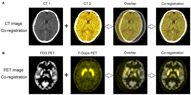
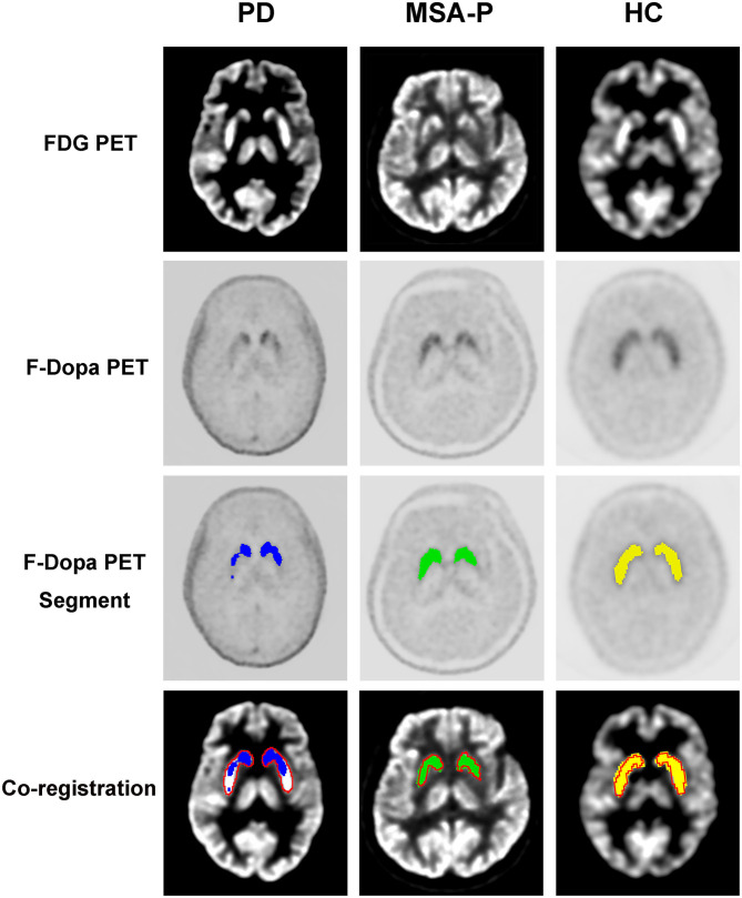
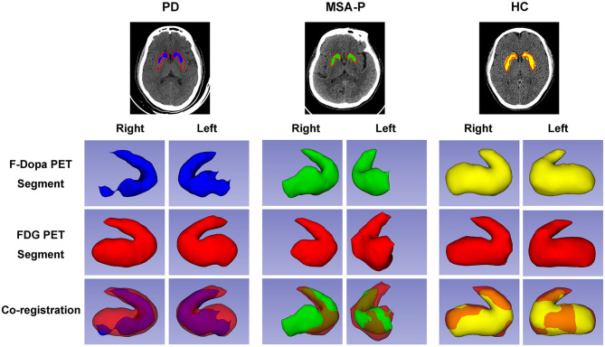
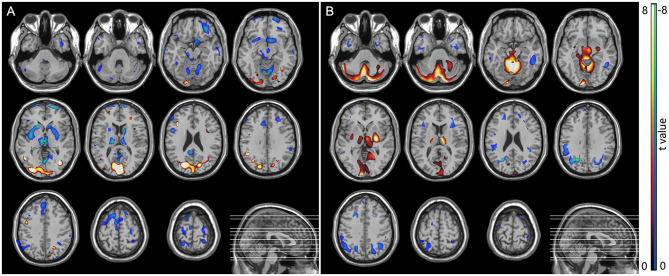
It is difficult to differentiate between Parkinson's disease and multiple system atrophy parkinsonian subtype (MSA-P) because of the overlap of their signs and symptoms. Enormous efforts have been made to develop positron emission tomography (PET) imaging to differentiate these diseases. This study aimed to investigate the co-registration analysis of 18F-fluorodopa and 18F-flurodeoxyglucose PET images to visualize the difference between Parkinson's disease and MSA-P. We enrolled 29 Parkinson's disease patients, 28 MSA-P patients, and 10 healthy controls, who underwent both 18F-fluorodopa and 18F-flurodeoxyglucose PET scans. Patients with Parkinson's disease and MSA-P exhibited reduced bilateral striatal 18F-fluorodopa uptake (p < 0.05, vs. healthy controls). Both regional specific uptake ratio analysis and statistical parametric mapping analysis of 18F-flurodeoxyglucose PET revealed hypometabolism in the bilateral putamen of MSA-P patients and hypermetabolism in the bilateral putamen of Parkinson's disease patients. There was a significant positive correlation between 18F-flurodeoxyglucose uptake and 18F-fluorodopa uptake in the contralateral posterior putamen of MSA-P patients (rs = 0.558, p = 0.002). Both 18F-flurodeoxyglucose and 18F-fluorodopa PET images showed that the striatum was rabbit-shaped in the healthy control group segmentation analysis. A defective rabbit-shaped striatum was observed in the 18F-fluorodopa PET image of patients with Parkinson's disease and MSA-P. In the segmentation analysis of 18F-flurodeoxyglucose PET image, an intact rabbit-shaped striatum was observed in Parkinson's disease patients, whereas a defective rabbit-shaped striatum was observed in MSA-P patients. These findings suggest that there were significant differences in the co-registration analysis of 18F-flurodeoxyglucose and 18F-fluorodopa PET images, which could be used in the individual analysis to differentiate Parkinson's disease from MSA-P.
Keywords: F-DOPA; FDG; PET; Parkinson's disease; mutiple system atrophy.
Copyright © 2021 Xian, Shi, Luo, Yi, Zhang and Pei.
Conflict of interest statement
The authors declare that the research was conducted in the absence of any commercial or financial relationships that could be construed as a potential conflict of interest.
Figures
Similar articles
-
The utility of susceptibility-weighted imaging for differentiating Parkinsonism-predominant multiple system atrophy from Parkinson's disease: correlation with 18F-flurodeoxyglucose positron-emission tomography.Neurosci Lett. 2015 Jan 1;584:296-301. doi: 10.1016/j.neulet.2014.10.046. Epub 2014 Nov 4. Neurosci Lett. 2015. PMID: 25450142
-
Separating Parkinson's disease from normality. Discriminant function analysis of fluorodopa F 18 positron emission tomography data.Arch Neurol. 1994 Mar;51(3):237-43. doi: 10.1001/archneur.1994.00540150027011. Arch Neurol. 1994. PMID: 8129633
-
Differential diagnosis of Parkinson's disease, multiple system atrophy, and Steele-Richardson-Olszewski syndrome: discriminant analysis of striatal 18F-dopa PET data.J Neurol Neurosurg Psychiatry. 1994 Mar;57(3):278-84. doi: 10.1136/jnnp.57.3.278. J Neurol Neurosurg Psychiatry. 1994. PMID: 8158173 Free PMC article.
-
Molecular imaging of movement disorders.World J Radiol. 2016 Mar 28;8(3):226-39. doi: 10.4329/wjr.v8.i3.226. World J Radiol. 2016. PMID: 27029029 Free PMC article. Review.
-
Voxelwise meta-analysis of gray matter anomalies in Parkinson variant of multiple system atrophy and Parkinson's disease using anatomic likelihood estimation.Neurosci Lett. 2015 Feb 5;587:79-86. doi: 10.1016/j.neulet.2014.12.007. Epub 2014 Dec 5. Neurosci Lett. 2015. PMID: 25484255
Cited by
-
Brain Evaluation by Dual PET/CT with [18F] FDOPA and [18F] FDG in Differential Diagnosis of Parkinsonian Syndromes.Brain Sci. 2024 Sep 18;14(9):930. doi: 10.3390/brainsci14090930. Brain Sci. 2024. PMID: 39335427 Free PMC article.
-
Identification of Parkinson's disease and multiple system atrophy using multimodal PET/MRI radiomics.Eur Radiol. 2024 Jan;34(1):662-672. doi: 10.1007/s00330-023-10003-9. Epub 2023 Aug 3. Eur Radiol. 2024. PMID: 37535155
-
Effects of STN-DBS surgery on cerebral glucose metabolism and distribution of DAT in Parkinson's disease.Brain Behav. 2023 Aug;13(8):e3172. doi: 10.1002/brb3.3172. Epub 2023 Jul 17. Brain Behav. 2023. PMID: 37459244 Free PMC article.
-
Involvement of striatal motoric subregions in familial frontotemporal dementia with parkinsonism harboring the C9orf72 repeat expansions.NPJ Parkinsons Dis. 2022 Oct 6;8(1):128. doi: 10.1038/s41531-022-00398-5. NPJ Parkinsons Dis. 2022. PMID: 36202819 Free PMC article.
-
Asymmetry in Atypical Parkinsonian Syndromes-A Review.J Clin Med. 2024 Sep 28;13(19):5798. doi: 10.3390/jcm13195798. J Clin Med. 2024. PMID: 39407856 Free PMC article. Review.
References
-
- Burn D. J., Sawle G. V., Brooks D. J. (1994). Differential diagnosis of Parkinson's disease, multiple system atrophy, and Steele-Richardson-Olszewski syndrome: discriminant analysis of striatal 18F-dopa PET data. J. Neurol. Neurosurg. Psychiatry 57, 278–284. 10.1136/jnnp.57.3.278 - DOI - PMC - PubMed
LinkOut - more resources
Full Text Sources
Other Literature Sources