. 2021 Apr 17:2021:4681041.
doi: 10.1155/2021/4681041. eCollection 2021.

Doxycycline Hyclate Modulates Antioxidant Defenses, Matrix Metalloproteinases, and COX-2 Activity Accelerating Skin Wound Healing by Secondary Intention in Rats

Affiliations

Doxycycline Hyclate Modulates Antioxidant Defenses, Matrix Metalloproteinases, and COX-2 Activity Accelerating Skin Wound Healing by Secondary Intention in Rats

Luciana S Altoé et al. Oxid Med Cell Longev. .

Abstract

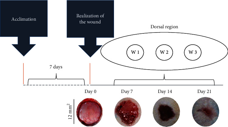
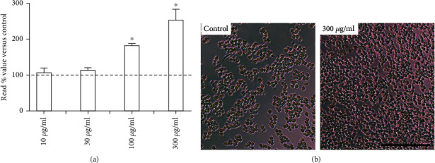
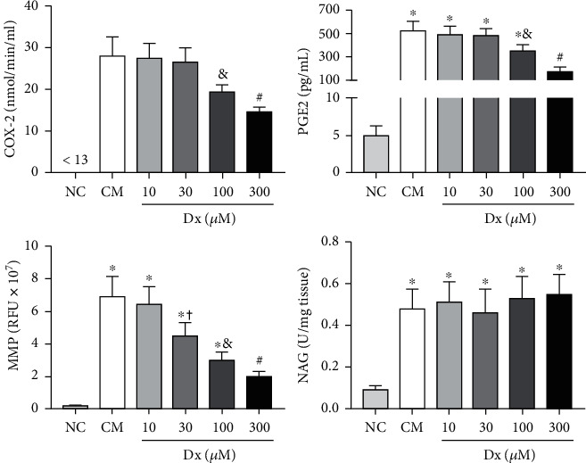
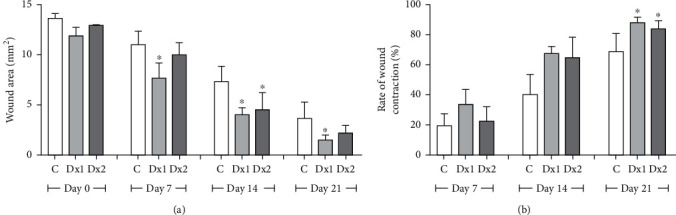
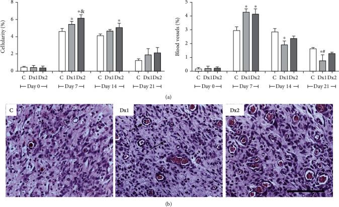
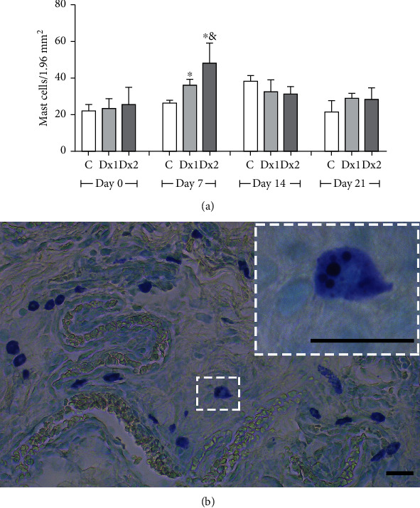
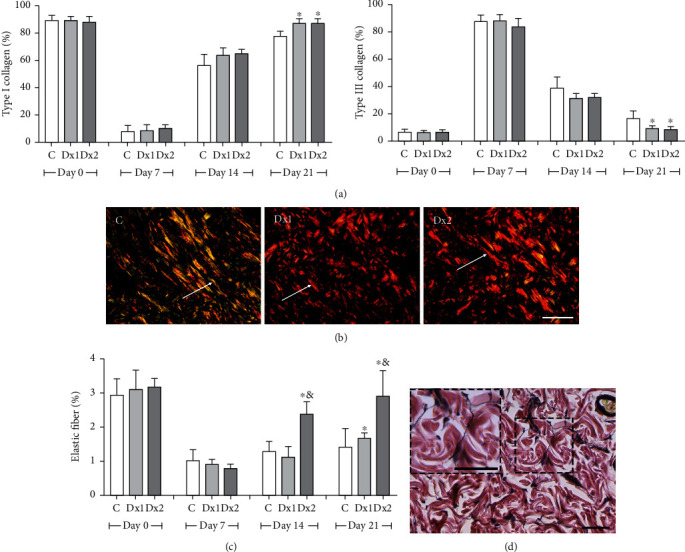
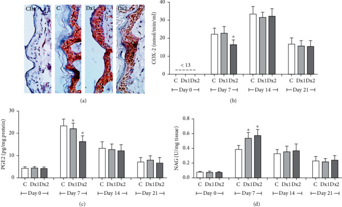
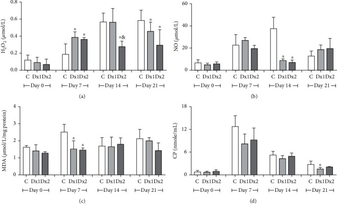
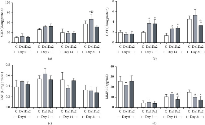
The main objective of this study was to investigate the action of doxycycline hyclate (Dx) in the skin wound healing process in Wistar rats. We investigated the effect of Dx on inflammatory cell recruitment and production of inflammatory mediators via in vitro and in vivo analysis. In addition, we analyzed neovascularization, extracellular matrix deposition, and antioxidant potential of Dx on cutaneous repair in Wistar rats. Male animals (n = 15) were divided into three groups with five animals each (protocol: 72/2017), and three skin wounds (12 mm diameter) were created on the back of the animals. The groups were as follows: C, received distilled water (control); Dx1, doxycycline hyclate (10 mg/kg/day); and Dx2, doxycycline hyclate (30 mg/kg/day). The applications were carried out daily for up to 21 days, and tissues from different wounds were removed every 7 days. Our in vitro analysis demonstrated that Dx led to macrophage proliferation and increased N-acetyl-β-D-glucosaminidase (NAG) production, besides decreased cyclooxygenase-2 (COX-2), prostaglandin E2 (PGE2), and metalloproteinases (MMP), which indicates that macrophage activation and COX-2 inhibition are possibly regulated by independent mechanisms. In vivo, our findings presented increased cellularity, blood vessels, and the number of mast cells. However, downregulation was observed in the COX-2 and PGE2 expression, which was limited to epidermal cells. Our results also showed that the downregulation of this pathway benefits the oxidative balance by reducing protein carbonyls, malondialdehyde, nitric oxide, and hydrogen peroxide (H2O2). In addition, there was an increase in the antioxidant enzymes (catalase and superoxide dismutase) after Dx exposure, which demonstrates its antioxidant potential. Finally, Dx increased the number of types I collagen and elastic fibers and reduced the levels of MMP, thus accelerating the closure of skin wounds. Our findings indicated that both doses of Dx can modulate the skin repair process, but the best effects were observed after exposure to the highest dose.


Conflict of interest statement

The authors declare that there are no conflicts of interest regarding the publication of this manuscript.

Figures

Figure 1
Figure 1
Representation of the experimental model of wound healing by secondary intention and time-dependent evolution of wound closure. The top image shows the distribution of the three excisional wounds in the back of the animal. The general appearance of wound closure from the initial wound (day 0) is represented by photographs. W1 (day 7), W2 (day 14), and W3 (day 21); macroscopic aspect of the wounds observed every 7 days. The wound areas were calculated on days 0, 7, 14, and 21 (mean ± SD), based on the digitized images.
Figure 2
Figure 2
Effects of doxycycline hyclate (Dx) on cell viability. (a) RAW264.7 macrophages were treated with various doses of Dx (10, 30, 100, and 300 μg/mL) for 24 h. (b) Representative photomicrographs showing cells in cultured medium (control group) and 300 μg/mL Dx added to the medium (phase-contrast microscopy, bar = 200 μm). The results are presented as the percentage absorbance of the control group. The data are expressed in the graphics as a mean and standard deviation. Statistical difference compared to control cells (Student-Newman-Keuls test, p < 0.05).
Figure 3
Figure 3
Effects of doxycycline hyclate (Dx) on cyclooxygenase-2 (COX-2), prostaglandin E2 production, and metalloproteinases (MMP) and N-acetyl-β-D-glucosaminidase (NAG) activity in LPS-stimulated RAW264.7 macrophages treated with various doses of Dx for 24 h. NC: not stimulated cells (not treated with LPS or Dx), CM: cells treated with culture medium containing LPS, and Dx: cells treated with culture medium containing LPS and Dx at 10, 30, 100, and 300 μM. The data are expressed as mean and standard deviation. Statistical difference (Student-Newman-Keuls test, p < 0.05), compared to NC, &CM, Dx10, and Dx30, #NC, CM, and Dx (10, 30, and 100), and + CM and Dx10.
Figure 4
Figure 4
(a) Area (mm2) and (b) rate of wound contraction (%) in rats treated with doxycycline hyclate (Dx) after 7, 14, and 21 days of treatment. Dx1: doxycycline hyclate (10 mg/kg), Dx2: doxycycline hyclate (30 mg/kg). The data are expressed as mean and standard deviation. Statistical difference (Kruskal–Wallis test, p < 0.05) compared to the control group.
Figure 5
Figure 5
The proportion of cell nucleus and blood vessels (a) and representative photomicrographs showing cells and blood vessel distribution (b) in the scar tissue of rats untreated and treated with doxycycline hyclate (Dx), on day 7 (H&E staining, bar = 100 μm). C: control, Dx1 = 10 mg/kg Dx, and Dx2 = 30 mg/kg Dx. In the graphics, the data are represented as mean and standard deviation. The statistical difference compared to the groups C, &Dx1, and #Dx2 (Student-Newman-Keuls test, p < 0.05).
Figure 6
Figure 6
The number of mast cells (a) and representative photomicrograph showing mast cell distribution (b) in the scar tissue from a rat treated with doxycycline hyclate (Dx, group 2) on day seven of wound healing (Toluidine blue staining, bar = 50 μm). C: control, Dx1 = 10 mg/kg Dx, and Dx2 = 30 mg/kg Dx. Data represented as mean and standard deviation. In the graphics, the data are represented as mean and standard deviation. The statistical difference compared to the groups C and &Dx1 (Student-Newman-Keuls test, p < 0.05).
Figure 7
Figure 7
The proportion of type I and III collagen fibers (a), representative photomicrographs showing collagen fiber distribution, on day 21 ((b) Sirius red staining under polarized light, bar = 100 μm), the proportion of elastic fibers (c), and representative photomicrograph showing elastic fiber distribution in group Dx2, on day 21 ((d) Verhoeff's elastic stain, bar = 50 μm) in the scar tissue of rats untreated and treated with doxycycline hyclate (Dx). C: control, Dx = 10 mg/kg Dx, and Dx2 = 30 mg/kg Dx. In the graphics, the data are represented as mean and standard deviation. The statistical difference compared to the groups C and &Dx1 (Student-Newman-Keuls test, p < 0.05).
Figure 8
Figure 8
Immunohistochemical detection of cyclooxygenase-2 (COX-2) in the epithelial cells (a), COX-2 activity (b), prostaglandin E2 (PGE2) levels (c), and N-acetyl-β-D-glucosaminidase (NAG) activity (d) in the scar tissue from rats untreated and treated with doxycycline hyclate (Dx). Cim: control of the immunohistochemical method (the primary antibody was omitted in the technique), C: control, Dx = 10 mg/kg Dx, and Dx2 = 30 mg/kg Dx. In the graphics, data are represented as mean and standard deviation. Statistical difference compared to group C (Student-Newman-Keuls test, p < 0.05).
Figure 9
Figure 9
Levels of oxidative stress markers in the tissue: (a) hydrogen peroxide (H2O2), (b) nitrite and nitrate (NO2/NO3), (c) malondialdehyde (MDA), and (d) carbonyl proteins (CP) in the scar tissue of rats untreated and treated with doxycycline hyclate (Dx). C: control, Dx1 = 10 mg/kg Dx, and Dx2 = 30 mg/kg Dx. Data are represented as mean and standard deviation. The statistical difference compared to the group C and &Dx1 (Student-Newman-Keuls test, p < 0.05).
Figure 10
Figure 10
Levels of (a) superoxide dismutase (SOD), (b) catalase (CAT), (c) glutathione S-transferase (GST), and (d) metalloproteinase-10 (MMP-10) in the scar tissue of rats untreated and treated with doxycycline hyclate (Dx). C: control, Dx1 = 10 mg/kg Dx, and Dx2 = 30 mg/kg Dx. The data are represented as mean and standard deviation. The statistical difference compared to the group C, &Dx1, and Dx2 (Student-Newman-Keuls test, p < 0.05).

References

    1. Marrot L. Pollution and sun exposure: a deleterious synergy. mechanisms and opportunities for skin protection. Current Medicinal Chemistry. 2019;25(40):5469–5486. doi: 10.2174/0929867324666170918123907. - DOI - PubMed
    1. Dai T., Huang Y., Sharma S. K., Hashmi J. T., Kurup D. B., Hamblin M. R. Topical antimicrobials for burn wound infections. Recent Patents on Anti-Infective Drug Discovery. 2010;5(2):124–151. doi: 10.2174/157489110791233522. - DOI - PMC - PubMed
    1. Badia J. M., Casey A. L., Petrosillo N., Hudson P. M., Mitchell S. A., Crosby C. Impact of surgical site infection on healthcare costs and patient outcomes: a systematic review in six European countries. Journal of Hospital Infection. 2017;96(1):1–15. doi: 10.1016/j.jhin.2017.03.004. - DOI - PubMed
    1. Sen C. K. Human wounds and its burden: an updated compendium of estimates. Advances in Wound Care. 2019;8(2):39–48. doi: 10.1089/wound.2019.0946. - DOI - PMC - PubMed
    1. Sarabahi S. Recent advances in topical wound care. Indian Journal of Plastic Surgery. 2012;45(2):379–387. doi: 10.4103/0970-0358.101321. - DOI - PMC - PubMed