Review
. 2021 Apr 20:8:637559.
doi: 10.3389/fmolb.2021.637559. eCollection 2021.

Virus Detection: A Review of the Current and Emerging Molecular and Immunological Methods

Affiliations
Review

Virus Detection: A Review of the Current and Emerging Molecular and Immunological Methods

A Cassedy et al. Front Mol Biosci. .

Abstract

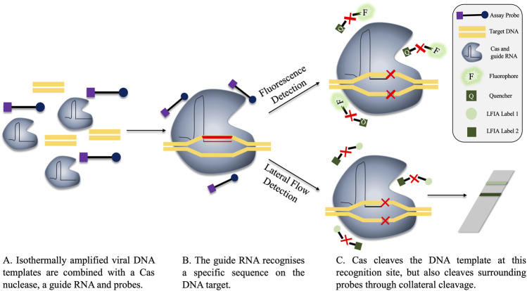
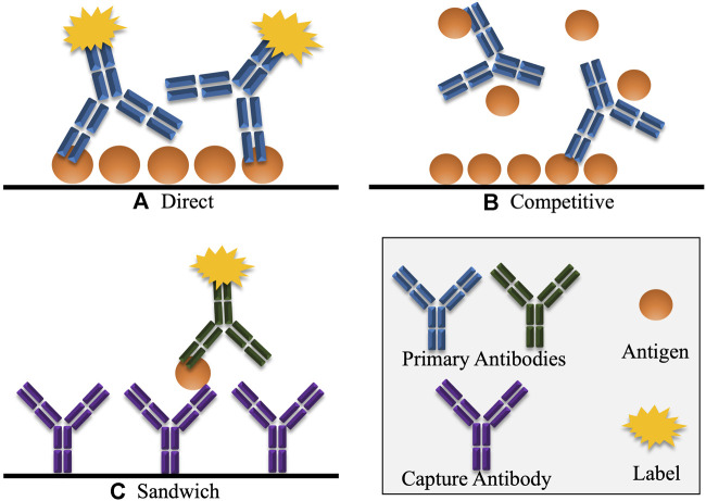
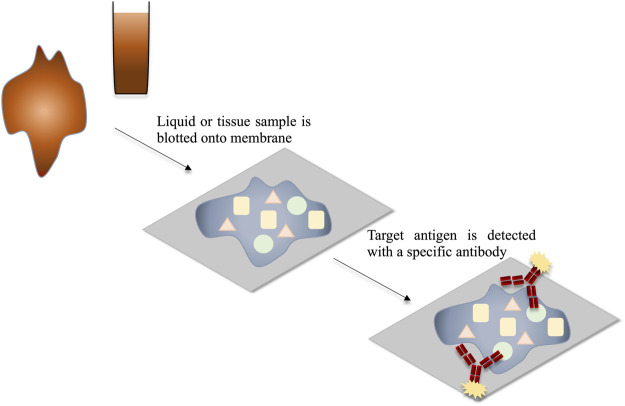
Viruses are ubiquitous in the environment. While many impart no deleterious effects on their hosts, several are major pathogens. This risk of pathogenicity, alongside the fact that many viruses can rapidly mutate highlights the need for suitable, rapid diagnostic measures. This review provides a critical analysis of widely used methods and examines their advantages and limitations. Currently, nucleic-acid detection and immunoassay methods are among the most popular means for quickly identifying viral infection directly from source. Nucleic acid-based detection generally offers high sensitivity, but can be time-consuming, costly, and require trained staff. The use of isothermal-based amplification systems for detection could aid in the reduction of results turnaround and equipment-associated costs, making them appealing for point-of-use applications, or when high volume/fast turnaround testing is required. Alternatively, immunoassays offer robustness and reduced costs. Furthermore, some immunoassay formats, such as those using lateral-flow technology, can generate results very rapidly. However, immunoassays typically cannot achieve comparable sensitivity to nucleic acid-based detection methods. Alongside these methods, the application of next-generation sequencing can provide highly specific results. In addition, the ability to sequence large numbers of viral genomes would provide researchers with enhanced information and assist in tracing infections.

Keywords: immunoassay; isothermal amplification; next generation sequencing; nucleic-acid detection; sampling; virus.


Conflict of interest statement

The authors declare that the research was conducted in the absence of any commercial or financial relationships that could be construed as a potential conflict of interest.

Figures

FIGURE 1
FIGURE 1
Comparison of CPE vs Pre-CPE detection in shell-vial culture. (A) Virus-containing sample is applied to the shell vial. (B) A centrifugal force is applied, encouraging inoculation of the virus into the cell monolayer. (C) The monolayer cells growing on a glass slide may be removed for interrogation. (D) CPE may take days to weeks to develop in the culturing cells. (E) Pre-CPE detection employs labelled antibodies to identify virus-specific markers; these can be detected within hours-days. (F) Microscopy can be used to inspect for morphological changes in CPE-diagnosis, or for signals arising from the presence of a labelled-antibody for pre-CPE detection.
FIGURE 2
FIGURE 2
The detection of target amplicon in qPCR through dyes and probes. The target region on the virus is amplified by multiple rounds of exponential amplification in PCR. During amplification, various methods are employed to monitor the real time production of target DNA. (A) Dyes intercalate indiscriminately with double stranded DNA, causing an increase in fluorescence as the level of double stranded DNA in the sample increases. Hydrolysis and hybridisation probes require binding to a specific sequence on the target amplicon to permit fluorescence. Fluorescence is achieved either by cleavage of the probe when employing hydrolysis probes (B), or through binding of the probes to the target region in close proximity to one another, as is the case for hybridisation probes (C).
FIGURE 3
FIGURE 3
Prevention of stem-loop formation caused by target sequence variations. (A) F1-F3, F1c-F3c, B1-B3 and B1c-B3c on the target DNA represent the binding regions for the LAMP primers, e.g. primers F3 and F2-F1c as shown in the diagram. If each of the primers matches suitably with the target DNA, stem loop formation proceeds. (B) Variation across any primer-binding sites (represented by yellow bands on target regions) can prevent stem-loop formation, and hence amplification. Primer design can be challenging when trying to avoid these variable regions.
FIGURE 4
FIGURE 4
A comparison of the various types of rolling circle amplification. (A) Linear amplification using a single primer. (B) Hyper-branched RCA adds a second primer which aids in exponential amplification by binding to regions on the repeated DNA sequence. (C) Multiply-primed RCA also facilitates the production of large amounts of amplicon via the use of several primers which bind to different regions on the circular DNA. (D) Padlocks probes contain sequences complementary to linear DNA. Hybridisation of these regions facilitates ligation, producing a circularised template which can be employed in RCA.
FIGURE 5
FIGURE 5
Fluorescence and lateral flow-based detection in a CRISPR-Cas detection system. (A) The components of the reaction are combined, either before or after isothermal amplification. The guide RNA is specific for a region on the target viral DNA. (B) The guide RNA recognises the target sequence, locating the Cas nuclease to this region. (C) The DNA target is cleaved by the Cas nuclease mechanism. However, Cas also performs collateral cleavage on surrounding single-stranded probes. Cleavage is represented in the diagram by a red X. These probes can be fluorescent (C, top), where the cleavage of the probe separates the quencher and the fluorophore, permitting fluorescent signal. Alternatively, probes can be used in lateral flow assays, whereby cleavage of the probe produces two distinct bands on the flow-strip, indicating a positive result. A single band on the lateral flow strip is representative of a negative result as no cleavage events occurred and the labels are not separated (C, bottom).
FIGURE 6
FIGURE 6
Various common formats of immunoassays, including ELISA. (A) In direct detection, the antigen is detected directly by a labelled antibody. Indirect detection is a variation of this format, where the primary antigen-specific antibody is unlabelled, and must be detected by a secondary labelled antibody that selectively binds to the primary antibody. (B) In competitive detection, free antigen in the sample competes with immobilised antigen for binding to the antibody which may be directly labelled or may be detected using a secondary labelled antibody. Hence, in competitive immunoassays, the signal is inversely proportional to the concentration of antigen. (C) In the sandwich immunoassay format, the antigen is captured by an antibody that reacts with a specific epitope on the antigen. A second labelled antibody is added which reacts with another different epitope on the captured antigen. Here the signal generated is directly proportional to the amount of antigen present.
FIGURE 7
FIGURE 7
Schematic representation of the detection of viral antigens through dot blot or tissue blot. The mixture of antigens within the sample is blotted onto the membrane and thereby immobilised. The target antigens are detected using target-specific labelled antibodies.

Similar articles

Cited by

References

    1. Adams I. P., Glover R. H., Monger W. A., Mumford R., Jackeviciene E., Navalinskiene M., et al. (2009). Next-generation sequencing and metagenomic analysis: a universal diagnostic tool in plant virology. Mol. Plant Pathol. 10, 537–545. 10.1111/j.1364-3703.2009.00545.x - DOI - PMC - PubMed
    1. Alexander T. S. (2016). Human immunodeficiency virus diagnostic testing: 30 Years of evolution. Clin. Vaccine Immunol. 23, 249–253. 10.1128/CVI.00053-16 - DOI - PMC - PubMed
    1. An L., Tang W., Ranalli T. A., Kim H. J., Wytiaz J., Kong H. (2005). Characterization of a thermostable UvrD helicase and its participation in helicase-dependent amplification. J. Biol. Chem. 280, 28952–28958. 10.1074/jbc.M503096200 - DOI - PMC - PubMed
    1. Arunrut N., Kampeera J., Sirithammajak S., Sanguanrut P., Proespraiwong P., Suebsing R., et al. (2016). Sensitive visual detection of AHPND bacteria using loop-mediated isothermal amplification combined with DNA-Functionalized gold nanoparticles as probes. PLoS One 11, e0151769. 10.1371/journal.pone.0151769 - DOI - PMC - PubMed
    1. Arya M., Shergill I. S., Williamson M., Gommersall L., Arya N., Patel H. R. (2005). Basic principles of real-time quantitative PCR. Expert Rev. Mol. Diagn. 5, 209–219. 10.1586/14737159.5.2.209 - DOI - PubMed

LinkOut - more resources