. 2021 Apr 20:8:639184.
doi: 10.3389/fmolb.2021.639184. eCollection 2021.

Enhanced siRNA Delivery and Selective Apoptosis Induction in H1299 Cancer Cells by Layer-by-Layer-Assembled Se Nanocomplexes: Toward More Efficient Cancer Therapy

Affiliations

Enhanced siRNA Delivery and Selective Apoptosis Induction in H1299 Cancer Cells by Layer-by-Layer-Assembled Se Nanocomplexes: Toward More Efficient Cancer Therapy

Maryam Sharifiaghdam et al. Front Mol Biosci. .

Abstract

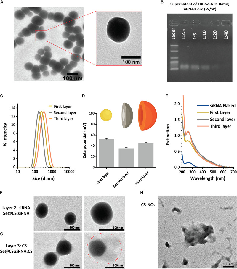
Nanotechnology has made an important contribution to oncology in recent years, especially for drug delivery. While many different nano-delivery systems have been suggested for cancer therapy, selenium nanoparticles (SeNPs) are particularly promising anticancer drug carriers as their core material offers interesting synergistic effects to cancer cells. Se compounds can exert cytotoxic effects by acting as pro-oxidants that alter cellular redox homeostasis, eventually leading to apoptosis induction in many kinds of cancer cells. Herein, we report on the design and synthesis of novel layer-by-layer Se-based nanocomplexes (LBL-Se-NCs) as carriers of small interfering RNA (siRNA) for combined gene silencing and apoptosis induction in cancer cells. The LBL-Se-NCs were prepared using a straightforward electrostatic assembly of siRNA and chitosan (CS) on the solid core of the SeNP. In this study, we started by investigating the colloidal stability and protection of the complexed siRNA. The results show that CS not only functioned as an anchoring layer for siRNA, but also provided colloidal stability for at least 20 days in different media when CS was applied as a third layer. The release study revealed that siRNA remained better associated with LBL-Se-NCs, with only a release of 35% after 7 days, as compared to CS-NCs with a siRNA release of 100% after 48 h, making the LBL nanocarrier an excellent candidate as an off-the-shelf formulation. When applied to H1299 cells, it was found that they can selectively induce around 32% apoptosis, while significantly less apoptosis (5.6%) was induced in NIH/3T3 normal cells. At the same time, they were capable of efficiently inducing siRNA downregulation (35%) without loss of activity 7 days post-synthesis. We conclude that LBL-Se-NCs are promising siRNA carriers with enhanced stability and with a dual mode of action against cancer cells.

Keywords: advanced drug delivery; apoptosis; cancer therapy; chitosan; nanomedicine; selenium nanoparticle; siRNA delivery.


Conflict of interest statement

The authors declare that the research was conducted in the absence of any commercial or financial relationships that could be construed as a potential conflict of interest.

Figures

SCHEME 1
SCHEME 1
Schematic illustration of layer-by-layer selenium nanocomplexes (LBL-Se-NCs). LBL-Se-NCs are composed of three layers: the core (Se@CS), a second layer with the cargo (siRNA), and a third protective layer of chitosan (CS). The nanocomplexes are taken up by cancer cells via endocytosis and trafficked via endosomes before finally escaping from endolysosomes. The released eGFP-siRNA and Se will induce a dual effect of apoptosis on the one hand and knockdown of the target protein on the other hand.
FIGURE 1
FIGURE 1
Synthesis of LBL-Se-NCs. (A) TEM images of Se@CS nanoparticles. The images confirmed the formation of SeNPs and indicate an average particle size of around 74.3 ± 5 nm. (B) The siRNA-loading capacity of Se@CS nanoparticles, from 1:2.5 to 1:40 siRNA:core mass ratios, was evaluated by measuring the remaining siRNA content in the supernatant by agarose gel electrophoresis. (C,D) The hydrodynamic diameter (C) and the surface charge (D) of the nanoparticles were evaluated with dynamic light scattering after each step in the synthesis process. The first layer corresponds to Se@CS, the second layer to Se@CS:siRNA, and the third layer to Se@CS:siRNA:CS. (n = 3). (E) UV-Visible spectra of the different stages of the LBL synthesis of Se-NCs. (F) TEM images of Se@CS:siRNA (G) and Se@CS:siRNA:CS. (H) TEM images of CS-NCs.
FIGURE 2
FIGURE 2
Evaluation of LBL-Se-NC colloidal stability and siRNA release. Characterization of LBL-Se-NCs and CS-NC (A) colloidal stability in different media: ddi. water (pH 7.4, 3, 9), DMEM, HEPES, PBS. (B) Colloidal stability as a function of time in RNase-free water as measured by DLS and zeta potential. (C) siRNA release profile from LBL-Se-NCs and CS-NCs as a function of time. Bars represent mean ± SEM, for a minimum of three independent experiments.
FIGURE 3
FIGURE 3
Comparison of biological effect by LBL-Se-NCs and CS-NCs in H1299 and NIH/3T3 cells. (A) Cell viability after treatment of H1299 (cancer cells) and NIH/3T3 (normal cells) with LBL-Se-NCs and CS-NCs. (B) Flow cytometric determination of apoptotic and necrotic populations of cells treated with LBL-Se-NCs in comparison with CS-NCs on H1299 and NIH/3T3 cells. (C) Uptake of AF647 siRNA-loaded Se-NCs and CS-NCs in H1299 and NIH/3T3 cell lines quantified via flow cytometry. ‘Uptake%’ refers to the percentage of cells that are positive for AF647. (D) The corresponding relative mean AF647 fluorescence intensity per cell (rMFI), which is proportional to the amount of particles that are taken up per cell on average. Bars represent mean ± SEM (n = 3, *p < 0.05; **p < 0.01; ***p < 0.001).
FIGURE 4
FIGURE 4
In vitro knockdown efficiency of LBL-Se-NCs. (A) GFP expression (%) of H1299 cells treated with LBL-Se-NCs as a function of siRNA concentration. (B) Comparison of the GFP expression level on H1299 cells after treatment with LBL-Se-NCs, Lipofectamine, jetPEI, CS-NCs, and naked siRNA, all at 8 nM effective siRNA concentration, for both freshly synthesized and 7-day-old nanocomplexes. (C) Confocal images of intracellular GFP knockdown of LBL-Se-NCs and control groups on H1299 cells. The scale bar corresponds to 50 μm. Data are presented as mean values ± SD (n = 3, *p < 0.05; **p < 0.01; ***p < 0.001).
FIGURE 5
FIGURE 5
Evaluation of Endosomal Escape based on AF647 ONs. (A) Representative confocal images 24 h post-incubation with AF647 ONs-loaded NCs in eGFP-H1299 cells. Upon endosomal escape, the labeled ONs which are loaded into the various nanocomplexes spread toward the cytoplasm, dequench, and finally accumulate into the nucleus. Hoechst nuclei can be seen in blue, while cells in which the endosomal escape occurred show nuclear fluorescence in the red channel (white arrows). The scale bar corresponds to 50 μm. (B) The histogram of red fluorescent intensity of cell nuclei. (C) Scheme of ONs uptake, endosomal escape, and accumulation in the nucleus. (D) The percentage of cells that had at least one endosomal escape event as quantified from AF647 fluorescence of at least 250 nuclei. The scale bar corresponds to 50 μm, (n = 3, *p < 0.05; **p < 0.01; ***p < 0.001).

Similar articles

Cited by

References

    1. Ahmad M. Z., Rizwanullah M., Ahmad J., Alasmary M. Y., Akhter M. H., Abdel-Wahab B. A., et al. (2021). Progress in nanomedicine-based drug delivery in designing of chitosan nanoparticles for cancer therapy. Int. J. Polym. Mater. 1–22
    1. Banjac A., Perisic T., Sato H., Seiler A., Bannai S., Weiss N., et al. (2008). The cystine/cysteine cycle: a redox cycle regulating susceptibility versus resistance to cell death. Oncogene 27 1618–1628. 10.1038/sj.onc.1210796 - DOI - PubMed
    1. Boroumand S., Safari M., Shaabani E., Shirza M., Faridi-Majidi R. (2019). Selenium nanoparticles: synthesis, characterization and study of their cytotoxicity, antioxidant and antibacterial activity. Mater. Res. Express 6:0850d0858.
    1. Buyens K., De Smedt S. C., Braeckmans K., Demeester J., Peeters L., van Grunsven L. A., et al. (2012). Liposome based systems for systemic siRNA delivery: stability in blood sets the requirements for optimal carrier design. J. Control. Release 158 362–370. 10.1016/j.jconrel.2011.10.009 - DOI - PubMed
    1. Buyens K., Lucas B., Raemdonck K., Braeckmans K., Vercammen J., Hendrix J., et al. (2008). A fast and sensitive method for measuring the integrity of siRNA-carrier complexes in full human serum. J. Control. Release 126 67–76. 10.1016/j.jconrel.2007.10.024 - DOI - PubMed

LinkOut - more resources