. 2021 May 28;478(10):1985-1997.
doi: 10.1042/BCJ20210017.

Insights into the substrate discrimination mechanisms of methyl-CpG-binding domain 4

Affiliations

Insights into the substrate discrimination mechanisms of methyl-CpG-binding domain 4

Hala Ouzon-Shubeita et al. Biochem J. .

Abstract

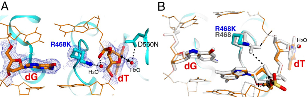
G:T mismatches, the major mispairs generated during DNA metabolism, are repaired in part by mismatch-specific DNA glycosylases such as methyl-CpG-binding domain 4 (MBD4) and thymine DNA glycosylase (TDG). Mismatch-specific DNA glycosylases must discriminate the mismatches against million-fold excess correct base pairs. MBD4 efficiently removes thymine opposite guanine but not opposite adenine. Previous studies have revealed that the substrate thymine is flipped out and enters the catalytic site of the enzyme, while the estranged guanine is stabilized by Arg468 of MBD4. To gain further insights into the mismatch discrimination mechanism of MBD4, we assessed the glycosylase activity of MBD4 toward various base pairs. In addition, we determined a crystal structure of MBD4 bound to T:O6-methylguanine-containing DNA, which suggests the O6 and N2 of purine and the O4 of pyrimidine are required to be a substrate for MBD4. To understand the role of the Arg468 finger in catalysis, we evaluated the glycosylase activity of MBD4 mutants, which revealed the guanidinium moiety of Arg468 may play an important role in catalysis. D560N/R468K MBD4 bound to T:G mismatched DNA shows that the side chain amine moiety of the Lys stabilizes the flipped-out thymine by a water-mediated phosphate pinching, while the backbone carbonyl oxygen of the Lys engages in hydrogen bonds with N2 of the estranged guanine. Comparison of various DNA glycosylase structures implies the guanidinium and amine moieties of Arg and Lys, respectively, may involve in discriminating between substrate mismatches and nonsubstrate base pairs.

Keywords: DNA glycosylase; base excision repair; substrate recognition.


Conflict of interest statement

Competing Interests: The authors declare no conflict of interest.

Figures

Figure 1.
Figure 1.. Base excision repair of G:T mismatches by MBD4.
(A) Formation and processing of G:T mismatches at CpG sites. DNMT: DNA methyltransferase; AID: activation induced cytidine deaminase; TDG: thymine DNA glycosylase; BER: base excision repair; TET2: Tet methylcytosine dioxygenase 2. (B) Extrahelical recognition of G:T mismatch by hMBD4. The flipped-out thymidine is recognized by Tyr540, Gln449 and Val448 and stabilized by Arg468-mediated phosphate pinching. The backbone carbonyl of Arg468 engages in hydrogen bonding interactions with the Watson-Crick edge of the estranged guanine. (C) Extrahelical recognition of G:T mismatch by MBD4.
Figure 2.
Figure 2.. The glycosylase activity of MBD4 toward purine:thymine base pairs.
(A) Base pairings of thymine with purines and purine analogs. O6MeG:T can form pseudo-Watson-Crick base pair. (B) Sequence of 27-bp DNA duplex used for the glycosylase activity assay. (C) Urea PAGE gel showing the glycosylase activity of MBD4 toward purine:T base pairs. Human MBD4 was incubated with dsDNA containing T paired with G, O6MeG, hypoxanthine (HX), 2,6-aminopurine (2,6-AP), or 2-aminopurine (2-AP) and the resulting reaction mixture was treated with sodium hydroxide. The sodium hydroxide-treated products were separated on a denaturing gel.
Figure 3.
Figure 3.. Single turnover kinetics of wild-type MBD4 and R468K MBD4 on G:T and/or O6MeG:T mismatches.
(A) DNA sequence used for kinetic studies. (B) Representative denaturing PAGE gel images for wild-type MBD4 and R468K mutant toward G:T- or O6MeG:T-containing DNA. (C) The activity of MBD4 catalytic domain on G:T and O6MeG mismatches under single turnover conditions ([EMBD4] = 2.5 μ;M, and [SDNA] = 0.25 μ;M) at pH 8.0 and room temperature).
Figure 4.
Figure 4.. Overall structure of D560N MBD4 in complex with O6MeG:T-containing DNA.
The substrate thymine opposite O6MeG lesion enters the catalytic site of MBD4 and the flipped-out thymine and O6MeG are stabilized by polar contacts with Arg468. Hydrogen bonds by Arg468 are indicated are dotted lines.
Figure 5.
Figure 5.. Structure of D560N MBD4 in complex with O6MeG:T-containing DNA.
(A) The active site of the D560N MBD4-O6MeG:T complex. A 2Fo-Fc electron density map contoured at 1σ around the substrate dT, estranged O6MedG, Arg468 and the catalytic water is shown. Note the electron density of the catalytic water is less defined compared to the catalytic water in the published D560N MBD4-G:T structure. (B) Superposition of D560N MBD4-O6MeG:T (cyan) and D560N MBD4-G:T (white) complexes. (C) Binding of D560N MBD4 to G:T- versus O6MeG:T-containing DNA. Electrophoretic mobility shift assay of D560N MBD4 with 50 nM of 27-bp-FAM-labeled G:T-containing DNA or O6MeG:T-containing DNA. DNA sequence used in this binding assay is shown in Figure 2B.
Figure 6.
Figure 6.. The Effect of Arg468 mutation on MBD4 glycosylase activity.
(A) The sequence of the FAM-labeled G:T-mismatch-containing duplex DNA used for the glycosylase activity assay. The G:T mismatch is indicated in red. (B) Glycosylase activity assay of MBD4 with G:T-containing DNA. Human MBD4, either wild type or Arg468 mutated as indicated, was incubated with dsDNA (shown above), treated with sodium hydroxide, and separated on denaturing gel. The cleaved DNA at the abasic site is labeled as product and travels faster than the longer uncleaved substrate.
Figure 7.
Figure 7.. Structure of R468K/D560N MBD4 in complex with G:T-containing DNA.
(A) Closer view of the active site of R468K/D560N MBD4-G:T complex. A 2FoFc electron density map contoured at 1σ around the substrate dT, estranged dG, Arg468, the catalytic water, and the water bridging the hydrogen bond interactions between R468K and the phosphate backbone of the base 3’ to the flipped dT is shown. (B) Superposition of the R468K/D560N MBD4-G:T (cyan) and D560N MBD4- G:T (white) complexes.
Figure 8.
Figure 8.. Representative intercalating amino acid residues observed in DNA glycosylase structures.
(A) hMBD4 (this work, 4OFA). Arg468 directly interacts with the 5’ phosphate oxygen of the substrate thymine (B) Bst MutY (3G0Q). Gln48 is hydrogen bonded to the 5’ phosphate oxygen of dA opposite 8-oxoguanine. (C) hAAG (1F4R). Tyr162 forms a water-mediated hydrogen bond with the 5’-phosphate oxygen of ethenoadenine opposite dT. (D) E. coli AlkA (1DIZ). Leu125 from the backbone carbonyl forms a water-mediated hydrogen bond with the 5’-phosphate of the abasic site analog. (E) hOGG1 (1EBM). Asn149 engages in a water-mediated hydrogen bond to the 5’-phosphate oxygen of 8-oxoguanine opposite dC. (F) hTDG (3UFJ). Arg275 forms a hydrogen bond with the 5’-phosphate oxygen of dU opposite dG.

References

    1. Arana ME, and Kunkel TA (2010) Mutator phenotypes due to DNA replication infidelity. Semin Cancer Biol 20, 304–311 - PMC - PubMed
    1. Kunkel TA (1985) The mutational specificity of DNA polymerase-beta during in vitro DNA synthesis. Production of frameshift, base substitution, and deletion mutations. J Biol Chem 260, 5787–5796 - PubMed
    1. Watt DL, Buckland RJ, Lujan SA, Kunkel TA, and Chabes A. (2016) Genome-wide analysis of the specificity and mechanisms of replication infidelity driven by imbalanced dNTP pools. Nucleic Acids Res 44, 1669–1680 - PMC - PubMed
    1. Koag MC, Nam K, and Lee S. (2014) The spontaneous replication error and the mismatch discrimination mechanisms of human DNA polymerase beta. Nucleic Acids Res 42, 11233–11245 - PMC - PubMed
    1. Wang W, Hellinga HW, and Beese LS (2011) Structural evidence for the rare tautomer hypothesis of spontaneous mutagenesis. Proc Natl Acad Sci U S A 108, 17644–17648 - PMC - PubMed

Publication types

LinkOut - more resources