

Differences in Specific Mass Density Between Dinoflagellate Life Stages and Relevance to Accumulation by Hydrodynamic Processes
- PMID: 33960400
- PMCID: PMC8596432
- DOI: 10.1111/jpy.13181
Differences in Specific Mass Density Between Dinoflagellate Life Stages and Relevance to Accumulation by Hydrodynamic Processes
Abstract
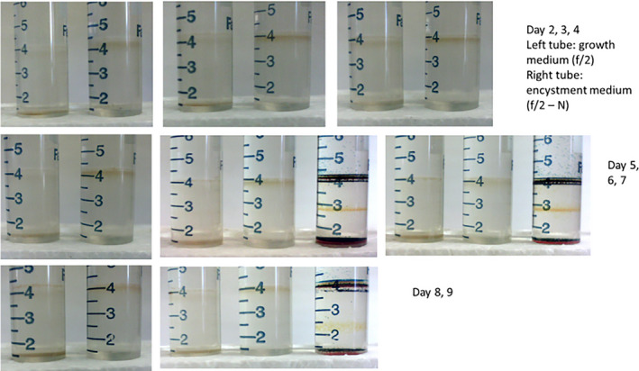
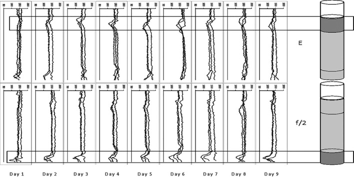
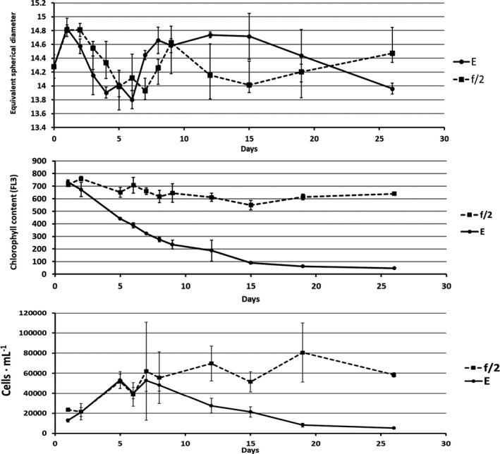
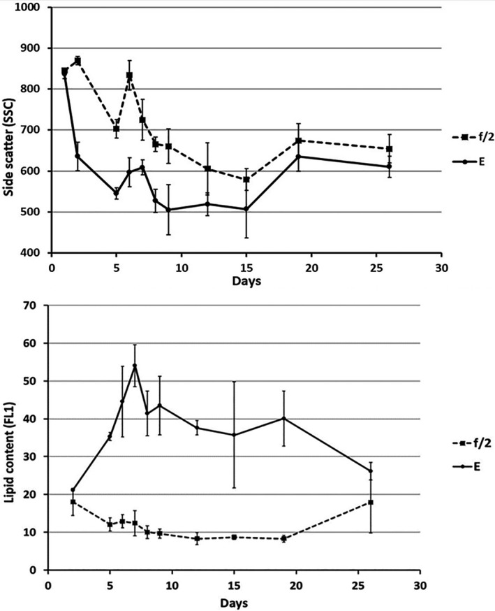
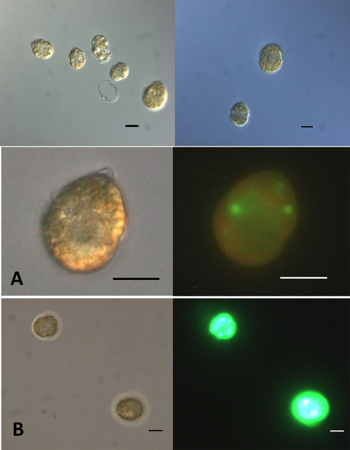
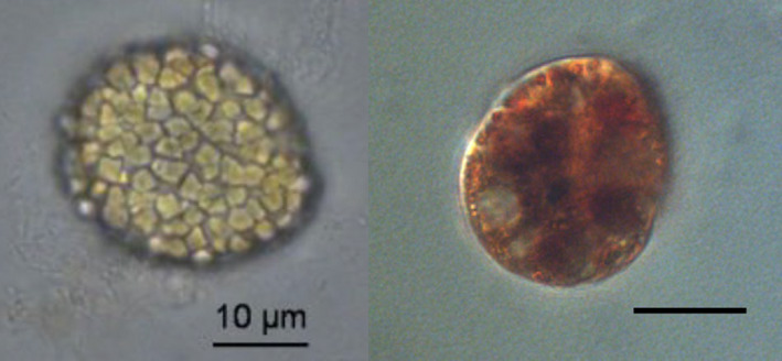
One previously unstudied aspect of differences between sexual and asexual life stages in large-scale transport and accumulation is density (mass per unit volume) of cells in each life stage. The specific density was determined for Scrippsiella lachrymosa cells in medium with and without nitrogen (N) enrichment through density-gradient centrifugation. Growth medium without N addition is often called "encystment medium" when used for the purpose of resting cyst formation in cyst-forming dinoflagellates; mating gametes are usually seen after 2-3 days. Significant differences in specific density were found after 2 days in encystment medium simultaneously with the observation of typical gamete swimming behavior and mating. The specific density of cells in encystment medium was 1.06 g · cm-3 ; whereas, the specific density of cells in growth medium was 1.11 g · cm-3 . Cells in encystment medium were found to have significantly increased lipid content, reduced chlorophyll content, and reduced internal complexity. The findings may explain differential transport of less dense and chemotactically aggregating gametes into surface blooms in contrast to denser vegetative cells that perform daily vertical migration and do not aggregate. Passive accumulation of non-migrating gametes into layers in stagnant water also can be explained, as well as sinking of zygotes when the storage of highly dense starch increases. Resting cysts had a density of over 1.14 g · cm-3 and would sink to become part of the silt fraction of the sediment. We suggest that differences in behavior and buoyancy between sexual and asexual life stages cause differences in cell accumulation, and therefore large-scale, environmental transport could be directly dependent upon life-cycle transitions.
Keywords: Scrippsiella lachrymosa; density gradient; dinoflagellate; encystment; gamete; percoll; sexual life stage.
© 2021 The Authors. Journal of Phycology published by Wiley Periodicals LLC on behalf of Phycological Society of America. This article is a U.S. Government work and is in the public domain in the USA.
Figures
Similar articles
-
Differences in pigmentation between life cycle stages in Scrippsiella lachrymosa (dinophyceae).J Phycol. 2016 Feb;52(1):64-74. doi: 10.1111/jpy.12364. Epub 2015 Dec 7. J Phycol. 2016. PMID: 26987089
-
Deficiency of nitrogen but not phosphorus triggers the life cycle transition of the dinoflagellate Scrippsiella acuminata from vegetative growth to resting cyst formation.Harmful Algae. 2022 Oct;118:102312. doi: 10.1016/j.hal.2022.102312. Epub 2022 Aug 26. Harmful Algae. 2022. PMID: 36195426
-
The life history of the toxic marine dinoflagellate Protoceratium reticulatum (Gonyaulacales) in culture.Harmful Algae. 2017 Sep;68:67-81. doi: 10.1016/j.hal.2017.07.008. Epub 2017 Aug 1. Harmful Algae. 2017. PMID: 28962991
-
Cyst-forming dinoflagellates in a warming climate.Harmful Algae. 2020 Jan;91:101728. doi: 10.1016/j.hal.2019.101728. Epub 2019 Dec 20. Harmful Algae. 2020. PMID: 32057345 Free PMC article. Review.
-
Trophic controls on stage transformations of a toxic ambush-predator dinoflagellate.J Eukaryot Microbiol. 1997 May-Jun;44(3):200-5. doi: 10.1111/j.1550-7408.1997.tb05700.x. J Eukaryot Microbiol. 1997. PMID: 9183706 Review.
Cited by
-
Marine Bioluminescence: Simulation of Dynamics within a Pump-Through Bathyphotometer.Sensors (Basel). 2024 Mar 19;24(6):1958. doi: 10.3390/s24061958. Sensors (Basel). 2024. PMID: 38544222 Free PMC article.
References
-
- Anderson, D. M. , Couture, D. A. , Kleindinst, J. L. , Keafer, B. A. , McGillicuddy, D. J. , Martin, J. L. , Richlen, M. L. , Hickey, J. M. & Solowa, A. R. 2014. Understanding interannual, decadal level variability in paralytic shellfish poisoning toxicity in the Gulf of Maine: the HAB Index. Deep‐Sea Res. II 103:264–76. - PMC - PubMed
-
- Anderson, D. M. , Keafer, B. A. , Kleindinst, J. L. , McGillicuddy, D. J. , Martin, J. L. , Norton, K. , Pilskaln, C. H. , Smith, J. L. , Sherwood, C. R. & Butman, B. 2014. Alexandrium fundyense cysts in the Gulf of Maine: long‐term time series of abundance and distribution, and linkages to past and future blooms. Deep. Res. II Top. Stud. Oceanogr. 103:6–26. - PMC - PubMed
-
- Anderson, D. M. , Lively, J. J. , Reardon, E. M. & Price, C. A. 1985. Sinking characteristics of dinoflagellate cysts. Limnol. Oceanogr. 30:1000–9.
-
- BD Biosciences 2000. Introduction to Flow Cytometry: A Learning Guide. San Jose, CA, 52 pp.
MeSH terms
Substances
LinkOut - more resources
Full Text Sources
Other Literature Sources
