Review
. 2021 Nov;83(11):e23254.
doi: 10.1002/ajp.23254. Epub 2021 May 7.

Studies of aging nonhuman primates illuminate the etiology of early-stage Alzheimer's-like neuropathology: An evolutionary perspective

Affiliations
Review

Studies of aging nonhuman primates illuminate the etiology of early-stage Alzheimer's-like neuropathology: An evolutionary perspective

Amy F T Arnsten et al. Am J Primatol. 2021 Nov.

Abstract

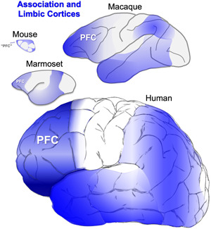
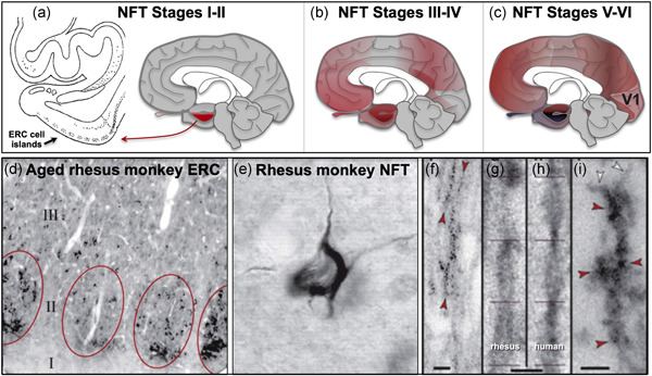
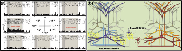
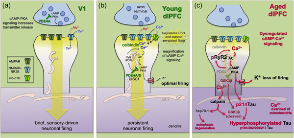
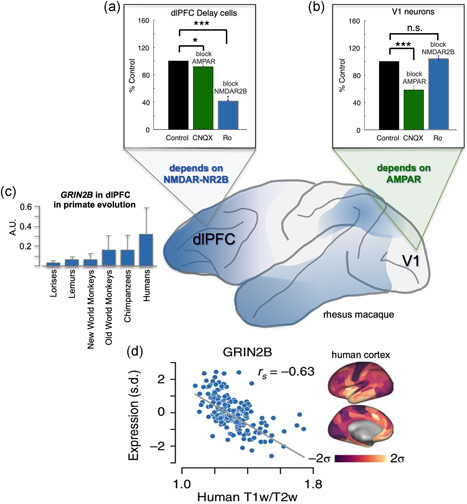
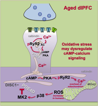
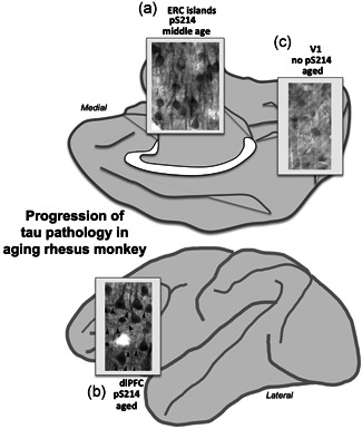
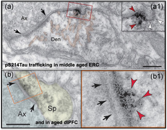
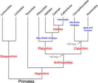
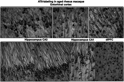
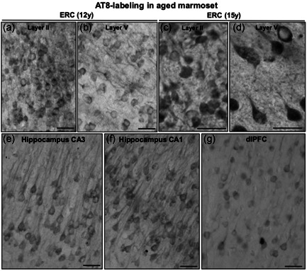
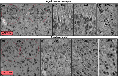
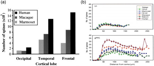
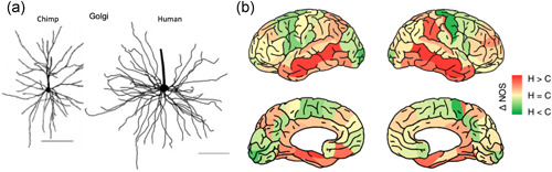
Tau pathology in Alzheimer's disease (AD) preferentially afflicts the limbic and recently enlarged association cortices, causing a progression of mnemonic and cognitive deficits. Although genetic mouse models have helped reveal mechanisms underlying the rare, autosomal-dominant forms of AD, the etiology of the more common, sporadic form of AD remains unknown, and is challenging to study in mice due to their limited association cortex and lifespan. It is also difficult to study in human brains, as early-stage tau phosphorylation can degrade postmortem. In contrast, rhesus monkeys have extensive association cortices, are long-lived, and can undergo perfusion fixation to capture early-stage tau phosphorylation in situ. Most importantly, rhesus monkeys naturally develop amyloid plaques, neurofibrillary tangles comprised of hyperphosphorylated tau, synaptic loss, and cognitive deficits with advancing age, and thus can be used to identify the early molecular events that initiate and propel neuropathology in the aging association cortices. Studies to date suggest that the particular molecular signaling events needed for higher cognition-for example, high levels of calcium to maintain persistent neuronal firing- lead to tau phosphorylation and inflammation when dysregulated with advancing age. The expression of NMDAR-NR2B (GluN2B)-the subunit that fluxes high levels of calcium-increases over the cortical hierarchy and with the expansion of association cortex in primate evolution, consistent with patterns of tau pathology. In the rhesus monkey dorsolateral prefrontal cortex, spines contain NMDAR-NR2B and the molecular machinery to magnify internal calcium release near the synapse, as well as phosphodiesterases, mGluR3, and calbindin to regulate calcium signaling. Loss of regulation with inflammation and/or aging appears to be a key factor in initiating tau pathology. The vast expansion in the numbers of these synapses over primate evolution is consistent with the degree of tau pathology seen across species: marmoset < rhesus monkey < chimpanzee < human, culminating in the vast neurodegeneration seen in humans with AD.

Keywords: AT8; Alzheimer's disease; calcium; entorhinal cortex; p217Tau; prefrontal cortex.


Figures

Figure 1
Figure 1
The association and limbic cortices (highlit in blue) expand across brain evolution
Figure 2
Figure 2
Tau pathology in humans and rhesus monkeys. (a) In human brain, cortical tau pathology begins in layer pre‐a in the perirhinal cortex and ERC (Braak stages I–II). The cell islands in layer II of the ERC are particularly prominent and indicated by a black arrow. (b) In Braak stages III–IV, tau pathology worsens in the ERC and medial temporal cortex, and builds in pyramidal cells of the association cortices. (c) Tau pathology only begins in the primary sensory cortices only at the latest stages, Braak stages V–VI. (d) Similar to humans, cortical tau pathology in aged rhesus monkeys begins in the layer II cell islands of the ERC. (e) Although not as frequent as in humans or chimpanzees, classic NFTs labeled by the AT8 antibody can be seen in the ERC of the oldest rhesus monkeys. (f–i) NFTs and pretangles in the aged rhesus monkey are comprised of paired helical filaments that label with the AT8 antibody used to diagnose AD (f), with the same helical frequency and size as human (g, h), ending abruptly as seen in human (i). ERC, entorhinal cortex; NFT, neurofibrillary tangle. (a–c) based on Arnsten et al. (2021) with permission; (d–i) from Paspalas et al. (2018) with permission
Figure 3
Figure 3
The cellular basis of working memory in the rhesus monkey dlPFC. (a) A “Delay cell” from the dlPFC of a rhesus monkey performing a visuospatial working memory task. Delay cells exhibit spatially tuned, persistent firing across the delay period when the monkey is remembering the spatial location that is the “preferred direction” for the neuron, but do not increase firing for nonpreferred directions. Thus, Delay cells are able to generate and sustain mental representations of visual space without sensory stimulation. (b) The recurrent excitatory microcircuits in deep layer III of rhesus monkey dlPFC are thought to underlie Delay cell firing. Pyramidal cells with a shared preferred direction excite each other through NMDAR synapses on spines to maintain firing across the delay period, while lateral inhibition from PV GABAergic interneurons helps to refine spatial tuning. dlPFC, dorsolateral prefrontal association cortex
Figure 4
Figure 4
Neurotransmission and neuromodulation in rhesus monkey V1 versus dlPFC, and its dysregulation with advancing age. (a) V1 neurons show classic characteristics, with neurotransmission relying heavily on AMPAR. In layer III of V1, PDE4A regulation of cAMP signaling is concentrated in presynaptic glutamatergic terminals, where cAMP‐PKA signaling can enhance glutamate release. Consistent with the immunoEM, physiological studies show that increasing cAMP‐PKA signaling in V1 increases neuronal firing. (b) Neurotransmission and neuromodulation are very different in layer III of the dlPFC, where neurotransmission relies heavily on NMDAR, including those with slowly closing NR2B subunits that flux high levels of calcium. PDE4 is concentrated in layer III spines on the SER, positioned to regulate cAMP‐PKA drive on internal calcium release, which in turn increases cAMP production, creating a vicious cycle that must be tightly regulated by PDE4s. While moderate levels of calcium are necessary to sustain persistent firing, higher levels of cAMP‐calcium signaling open nearby potassium (K+) channels to weaken connectivity and reduce firing, for example, as occurs with uncontrollable stress. In young adult dlPFC, layer III dlPFC pyramidal cells express high levels of PDE4s to regulate cAMP drive on calcium release, and calbindin to regulate calcium levels when it is released into the cytosol. These regulatory factors are lost with advanced age. (c) The loss of PDE4s and calbindin in aged dlPFC pyramidal cells leads to excessive calcium‐cAMP‐PKA signaling and initiates a series of vicious cycles and toxic events including: (1) PKA phosphorylation of RyR2 to cause calcium leak into the cytosol, leading to more cAMP‐PKA signaling; (2) excessive opening of K+ channels that reduce neuronal firing; (3) calcium overload of mitochondria to induce inflammatory signaling (see Figure 6); (4) PKA phosphorylation of tau, which primes tau for hyperphosphorylation by GSK3β; (5) with sufficient cytosolic calcium, the activation of calpain, which disinhibits GSK3β to hyperphosphorylate tau at key sites that lead to tau fibrillation; and (6) calpain activation of heatshock protein 70.1 to drive autophagic degeneration. dlPFC, dorsolateral prefrontal association cortex
Figure 5
Figure 5
NMDAR‐NR2B neurotransmission increases across the cortical hierarchy and across primate evolution. (a) Delay cells in rhesus monkey dlPFC rely on NMDAR‐NR2B neurotransmission, with relatively little influence from AMPAR. Thus, blockade of AMPAR with CNQX has little effect, while blockade of NMDAR with NR2B subunits with Ro25‐6981 markedly reduces Delay cell firing as the monkey performs a working memory task. (b) In contrast, neurons in rhesus monkey V1 depend on AMPAR more than NMDAR. Neurons responding to a bar of light greatly reduce their firing when AMPAR is blocked with CNQX, but show little effect when NMDAR‐NR2B is blocked by Ro25‐6981. (c) Increased expression of GRIN2B, which encodes for the NMDAR‐NR2B, in the dlPFC across primate evolution. (d) GRIN2B expression increases across the human cortical hierarchy. dlPFC, dorsolateral prefrontal association cortex. Panels A and B from Yang et al. (2018) with permission. Panel C from Muntané et al. (2015) with permission. Panel D from Burt et al. (2018) with permission.
Figure 6
Figure 6
Schematic illustration of the hypothesis that elevated ROS (oxidative stress) in dysmorphic mitochondria can induce p38‐MK2 signaling, which can dysregulate calcium signaling by unanchoring and inhibiting PDE4s, which in turn can create calcium overload of mitochondria, perpetuating a vicious cycle. Calcium dysregulation and p38 signaling can both contribute to the phosphorylation of tau. Higher rates of energy metabolism in human brain may increase these factors in humans, propelling AD pathology. MOAS, mitochondria‐on‐a‐string, an abnormal phenotype seen in AD and aged monkey association cortex; AD, Alzheimer's disease; ROS, reactive oxygen species
Figure 7
Figure 7
Early stage tau pathology (PKA phosphorylation of tau at pS214Tau) across the aging rhesus monkey cortex shows the same pattern and progression as in human, starting in the ERC layer II cell islands in middle age (a), and then with advanced age in the association cortices such as the dlPFC (b), but not in V1 even in very old animals (c). dlPFC, dorsolateral prefrontal association cortex. Image of tau pathology in A from Paspalas et al. (2018) with permission; images of tau pathology in B and C from Carlyle et al. (2014) with permission
Figure 8
Figure 8
pS214Tau trafficking between neurons near a glutamatergic synapse in layer II of the middle‐aged ERC (a), and within a glutamatergic synapse in the aged dlPFC (b). Enlarged to show details. dlPAC, dorsolateral prefrontal association cortex; ERC, entorhinal cortex. (a) from Paspalas et al. (2018) with permission; (b) from Carlyle et al. (2014) with permission
Figure 9
Figure 9
Evolutionary relationships among primates. Humans and apes comprised a higher‐order group, Hominoidea, which is related to another higher‐order group, Cercopithecoidea, which includes macaque monkeys and other Old World monkeys. Collectively, these are the Old World anthropoid primates, or the Catarrhini. The New World anthropoids, or Platyrrhini, include primates such as marmosets, squirrel monkeys, and capuchins. The species discussed in this review are highlighted in blue. Figure modified from Preuss (2019) with permission
Figure 10
Figure 10
Tau fibrillation (AT8 labeling) in aged rhesus macaque (31 years). Dense tau fibrillation is observed aggregating in entorhinal cortex layer II cell islands (a), extending into the deeper layers, including layer III (b) and layer V (c). The fibrillated tau‐labeling pattern is especially prominent in the perisomatic compartment and along apical and basilar dendrites (pretangles) of excitatory neurons, including parallel bundles of “twisting” apical dendrites in entorhinal cortex layer III and layer V, indicative of possible neurodegeneration. Prominent aggregated AT8 labeling is visualized in AD‐related vulnerable brain regions, including hippocampus pyramidal cell CA3 (d, e) and CA1 (f) subfields, and recurrent excitatory circuits in dlPFC layer III (g). Scale bars, 25 µm (a–c) and 30 µm (d–g). AD, Alzheimer's disease; dlPFC, dorsolateral prefrontal association cortex
Figure 11
Figure 11
Tau fibrillation (AT8 labeling) in aged marmoset (12–15 years). In early aged marmoset (12 years), mild AT8 labeling is observed in stellate cell layer II islands (a) and deeper layer V (b) in entorhinal cortex. The labeling pattern is diffuse and visualized in the cell soma and the proximal segment along apical dendrites. With more advanced age in marmosets (15 years), more fibrillated AT8 labeling is observed aggregating in entorhinal cortex layer II (c) and layer V (d). Mild tau fibrillation is visualized in pyramidal neurons in hippocampus CA3 (e) and CA1 (f) subfields, and dlPFC layer III (g) in aged marmoset. Scale bars, 25 µm
Figure 12
Figure 12
Multiregional pT217‐tau labeling in aged rhesus macaque and marmoset. In aged rhesus macaque (30–31 years), stellate cell layer II islands (red ovals) in entorhinal cortex (a), hippocampus CA3 (b), hippocampus CA1 (c), and dlPFC layer III (d) are reactive against pT217‐tau. Immunoreactivity for pT217‐tau is visualized in the cytoplasm of the perisomatic compartment, along apical and basilar dendrites, and within the nucleoplasm. Note, in aged rhesus macaque dlPFC (30 years), pT217‐tau labeling reveals a highly fibrillated pattern, reflected in “twisting” apical dendrites, similar to AT8. In aged marmoset (12 years), stellate cell layer II islands (red ovals) in entorhinal cortex (e), hippocampus CA3 (f), hippocampus CA1 (g), and dlPFC layer III (h) are reactive against pT217‐tau. Immunoreactivity for pT217‐tau in aged marmosets is more diffuse compared to rhesus macaques, but observed prominently in the neuronal soma, and proximal and distal segments of apical dendrites. Scale bars, 25 µm (a–c, e–g), 10 µm (d), 20 µm (h)
Figure 13
Figure 13
(a) Bar graph of the numbers of spines in V1 (occipital cortex), and the temporal and prefrontal association cortices from marmoset, macaque, and human, showing an evolutionary expansion across the cortical hierarchy and across primate evolution. From Elston et al. (2001) with permission. (b) The numbers of spines in V1 versus dlPFC across primate species, with no differences in V1, and a great expansion across species in dlPFC. dlPFC, dorsolateral prefrontal association cortex. From Elston (2006) with permission
Figure 14
Figure 14
(a) dlPFC pyramidal cell basal dendrites in the chimpanzee are less complex than those in human. From Bianchi et al. (2013) with permission. (b) Measures of connectivity determined from MRI assays of white matter in chimpanzees versus humans. Cortical regions with greater connectivity in humans are shown in red, those with greater connectivity in chimpanzees are shown in green. dlPFC, dorsolateral prefrontal association cortex; MRI, magnetic resonance imaging. From Ardesch et al. (2019) with permission

References

    1. Ardesch, D. J. , Scholtens, L. H. , Li, L. , Preuss, T. M. , Rilling, J. K. , & van den Heuvel, M. P. (2019). Evolutionary expansion of connectivity between multimodal association areas in the human brain compared with chimpanzees. Proceedings of the National Academy of Sciences of the United States of America, 116, 7101–7106. - PMC - PubMed
    1. Area‐Gomez, E. , & Schon, E. A. (2017). On the pathogenesis of Alzheimer's disease: The MAM Hypothesis. FASEB Journal, 31, 864–867. - PMC - PubMed
    1. Arnsten, A. F. (2015). Stress weakens prefrontal networks: Molecular insults to higher cognition. Nature Neuroscience, 18, 1376–1385. - PMC - PubMed
    1. Arnsten, A. F. T. , Datta, D. , Del Tredici, K. , & Braak, H. (2021). Hypothesis: Tau pathology is an initiating factor in sporadic Alzheimer's disease. Alzheimer's & Dementia, 17, 115–124. - PMC - PubMed
    1. Arnsten, A. F. T. , Datta, D. , & Wang, M. (2020). The genie in the bottle‐magnified calcium signaling in dorsolateral prefrontal cortex. Molecular Psychiatry. 10.1038/s41380-020-00973-3 - DOI - PMC - PubMed

Publication types

LinkOut - more resources