Development of angle closure and associated risk factors: The Handan eye study
- PMID: 33960669
- PMCID: PMC9292978
- DOI: 10.1111/aos.14887
Development of angle closure and associated risk factors: The Handan eye study
Abstract
Purpose: To investigate the development of angle closure from baseline open angle and associated risk factors in a rural Chinese population through a longitudinal study over a 5-year period.
Methods: Subjects aged ≥30 years and older with bilateral open angles at baseline of the Handan Eye Study who participated in the follow-up and had undergone both baseline and follow-up gonioscopic examinations were included. Subjects with any form of angle closure, glaucoma, incisional ocular surgery or other conditions that could influence the results were excluded. The development of angle closure was defined as the presence of primary angle closure suspect (PACS) or primary angle closure (PAC)/primary angle closure glaucoma (PACG) during the follow-up in normal subjects with baseline bilateral open angles. Logistic regression was performed to identify the baseline risk factors for the development of angle closure.
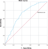
Results: A total of 457 subjects with bilateral open angles at baseline aged 53.0 (45.5, 58.0) years were enrolled. 94.7% of the included cases developed PACS, 5.3% developed PAC and no one developed PACG after 5 years. In logistic regression, significant risk factors for the development of angle closure were shallower central anterior chamber depth (ACD) (p = 0.002) and narrower mean angle width (p < 0.001).
Conclusions: This study reports the development from baseline open angle to angle closure after a 5-year follow-up. We confirm that the mean angle width and central ACD were independent predictive risk factors for the development of any form of angle closure.
Keywords: development of angle closure; primary angle closure; primary angle closure glaucoma; primary angle closure suspect; risk factors.
© 2021 The Authors. Acta Ophthalmologica published by John Wiley & Sons Ltd on behalf of Acta Ophthalmologica Scandinavica Foundation.
Figures
References
-
- Alsbirk PH (1992): Anatomical risk factors in primary angle‐closure glaucoma. A ten year follow up survey based on limbal and axial anterior chamber depths in a high risk population. Int Ophthalmol 16: 265–272. - PubMed
-
- Casson RJ, Baker M, Edussuriya K, Senaratne T, Selva D & Sennanayake S (2009): Prevalence and determinants of angle closure in central Sri Lanka: the Kandy Eye Study. Ophthalmology 116: 1444–1449. - PubMed
-
- Chylack LT Jr, Wolfe JK, Singer DM et al. (1993): The lens opacities classification system III. The longitudinal study of cataract study group. Arch Ophthalmol 111: 831–836. - PubMed
-
- Erie JC, Hodge DO & Gray DT (1997): The incidence of primary angle‐closure glaucoma in Olmsted County, Minnesota. Arch Ophthalmol 115: 177–181. - PubMed
Publication types
MeSH terms
Grants and funding
LinkOut - more resources
Full Text Sources
Other Literature Sources
Medical


