. 2021 May 7;19(5):e3001209.
doi: 10.1371/journal.pbio.3001209. eCollection 2021 May.

Structural basis for SARS-CoV-2 neutralizing antibodies with novel binding epitopes

Affiliations

Structural basis for SARS-CoV-2 neutralizing antibodies with novel binding epitopes

Dan Fu et al. PLoS Biol. .

Abstract

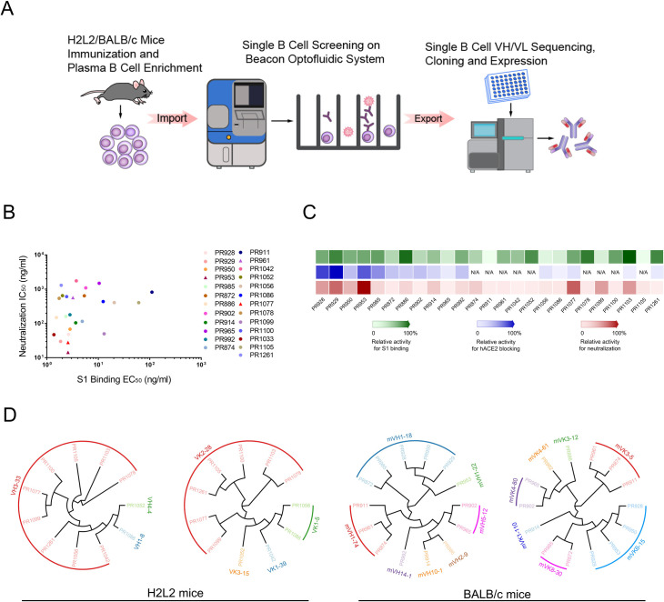
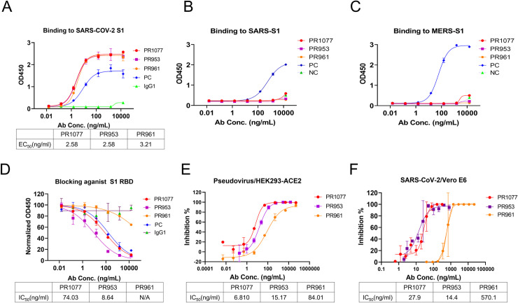
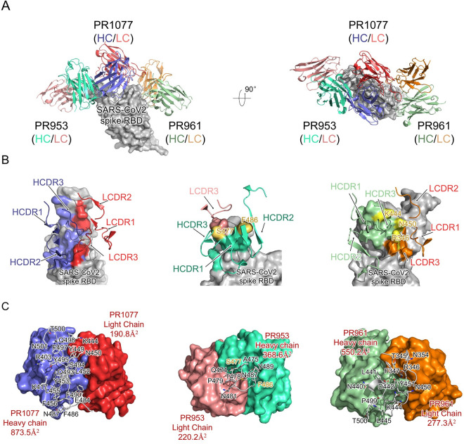
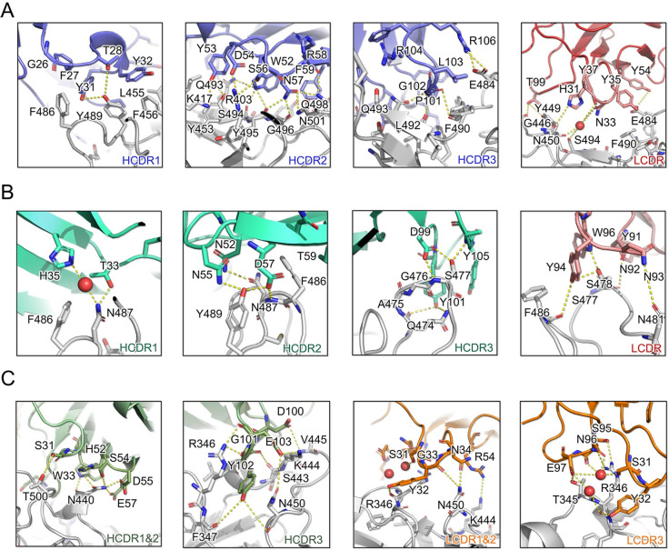
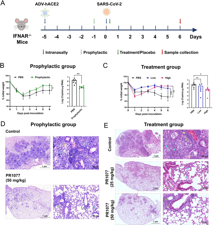
The ongoing Coronavirus Disease 2019 (COVID-19) pandemic caused by Severe Acute Respiratory Syndrome Coronavirus 2 (SARS-CoV-2) threatens global public health and economy unprecedentedly, requiring accelerating development of prophylactic and therapeutic interventions. Molecular understanding of neutralizing antibodies (NAbs) would greatly help advance the development of monoclonal antibody (mAb) therapy, as well as the design of next generation recombinant vaccines. Here, we applied H2L2 transgenic mice encoding the human immunoglobulin variable regions, together with a state-of-the-art antibody discovery platform to immunize and isolate NAbs. From a large panel of isolated antibodies, 25 antibodies showed potent neutralizing activities at sub-nanomolar levels by engaging the spike receptor-binding domain (RBD). Importantly, one human NAb, termed PR1077, from the H2L2 platform and 2 humanized NAb, including PR953 and PR961, were further characterized and subjected for subsequent structural analysis. High-resolution X-ray crystallography structures unveiled novel epitopes on the receptor-binding motif (RBM) for PR1077 and PR953, which directly compete with human angiotensin-converting enzyme 2 (hACE2) for binding, and a novel non-blocking epitope on the neighboring site near RBM for PR961. Moreover, we further tested the antiviral efficiency of PR1077 in the Ad5-hACE2 transduction mouse model of COVID-19. A single injection provided potent protection against SARS-CoV-2 infection in either prophylactic or treatment groups. Taken together, these results shed light on the development of mAb-related therapeutic interventions for COVID-19.


Conflict of interest statement

I have read the journal’s policy and the authors of this manuscript have the following competing interests: Y.G., Z.R., L.L., J.W., Z.L., D.F., X.W. and C.N. are inventors in a pending patent application filed on the reported antibodies. L.L., J.W., Z.L., X.W., C.N., C.D., L.Z., G.G. and N.S. are employees of Harbour Biomed (Suzhou) Co. Ltd. Other authors declare no competing interests. All reagents and information presented in this study are available from corresponding authors upon reasonable request.

Figures

Fig 1
Fig 1. Analysis of plasma responses to SARS-CoV-2 proteins and antibody identification from H2L2 transgenic and BALB/c mice by single cell sequencing.
(A) Schematic diagram of antibody identification from convalescent patients by single cell sequencing. Both Harbour H2L2 transgenic mice and BALB/c mice were immunized with SARS-CoV-2 RBD protein. SARS-CoV-2 RBD protein binding B cells were isolated from enriched mouse spleen and bone marrow cells with beads conjugated with biotinylated SARS-CoV-2 RBD protein. Paired VH and VL of each cell were recovered by single B cell sequencing. The sequences were used for subsequent antibody construction and expression. (B) Antibodies neutralization IC50(y axis) are plotted against SARS-CoV-2 S1 protein binding EC50 (x axis), PR1077, PR953, and PR961 are shown as triangle, and other antibodies are shown as dot. All the data of this figure can be found in the S1 Data file. (C) Heat map shows the relative levels of SARS-CoV-2 S1 binding, hACE2 blocking, and live virus neutralization. The depth of color is proportional to the effect of the antibody (green, S1 binding activity; blue, hACE2 blocking activity; red, neutralizing activity). All the data of this figure can be found in the S2 Data file. (D) Phylogenetic analysis of the relationship between antibody variable gene segments and germline. The relationships between the heavy and light chain variable regions and the germlines from H2L2 transgenic (left) and BALB/c (right) mouse antibodies are shown. hACE2, human angiotensin-converting enzyme 2; RBD, receptor-binding domain; SARS-CoV-2, Severe Acute Respiratory Syndrome Coronavirus 2.
Fig 2
Fig 2. Characterization of the selected NAbs PR1077, PR953, and PR961.
(A) ELISA binding curve of SARS-CoV-2 S1 protein. A commercially available antibody (8A5, Novoprotein, Shanghai) was applied as a PC. Normal human IgG1 was used as a negative control. All the data of this figure can be found in the S3 Data file. ELISA binding curves of SARS S1 (B) and MERS S1 (C) proteins. Internally generated antibodies were used as PCs. All the data of these figures can be found in the S4 Data and S5 Data files. (D) ELISA-based RBA of PR1077, PR953, and PR961 against RBD binding to hACE2. All the data of this figure can be found in the S6 Data file. (E) PR1077, PR953, and PR961 could effectively neutralize SARS-CoV-2 pseudovirus in vitro. SARS-CoV-2 pseudovirus was incubated with serially diluted antibodies. The mixture was added to HEK293-hACE2 cells for 48 hours. The neutralization potency of each antibody was evaluated in a luciferase assay system. All the data of this figure can be found in the S7 Data file. (F) PR1077, PR953, and PR961 showed strong neutralizing potency against live SARS-CoV-2 virus in vitro. The mixture of live SARS-CoV-2 virus and serially diluted PR1077 were added to Vero E6 cells. After 1 hour, cells were washed and further incubated for 48 hours before detection of infected cells by an immunofluorescence assay. All the data of this figure can be found in the S8 Data file. ELISA, enzyme-linked immunosorbent assay; NAb, neutralizing antibody; PC, positive control; RBA, receptor blocking assay; SARS-CoV-2, Severe Acute Respiratory Syndrome Coronavirus 2.
Fig 3
Fig 3. Structural analysis of the SARS-CoV-2 RBD-NAbs scFv complex.
(A) Overall structures of the PR1077, PR953, and PR961-Fab-RBD complexes. The PR1077 HC (colored slate blue) and light chain (colored red) are shown. PR953 heavy (colored green cyan) and light (colored salmon red) chains and PR961 heavy (colored pale green) and light (colored orange) chains are displayed. The SARS-CoV-2 RBD is colored in gray and displayed in surface representation. (B) The epitopes of antibodies are shown in surface representation. The CDR loops of NAbs are colored as above. The S477 and F486 residues in contact with both the heavy and light chains of PR953 are colored in yellow; 3 hydrogen donor residues (R346, K444, and N450) which contact with both the heavy and light chains of PR961 are colored in yellow. (C) The binding surface of NAbs with SARS-CoV-2-RBD. The interaction residues of RBD are shown in sticks and labeled accordingly; variable regions’ interaction surfaces of NAbs are colored as above. HC, heavy chain; HCDR, heavy chain complementarity-determining region; LCDR, light chain complementary-determining region; NAb, neutralizing antibody; RBD, receptor-binding domain; SARS-CoV-2, Severe Acute Respiratory Syndrome Coronavirus 2; scFv, single-chain variable fragment.
Fig 4
Fig 4. Interactions between the SARS-CoV-2 RBD and CDR loops.
(A–C) Detailed analysis of the interfaces of SARS-CoV-2 RBD with HCDR and LCDR for (A) PR1077, (B) PR953, and (C) PR961. For clarity, only the key residues are labeled: red, oxygen atoms; blue, nitrogen atoms; yellow dash lines, hydrogen bond interactions. The residues are shown in sticks with identical colors to Fig 3; water molecules are displayed as red spheres. CDR, complementary-determining region; HCDR, heavy chain complementarity-determining region; LCDR, light chain complementary-determining region; RBD, receptor-binding domain; SARS-CoV-2, Severe Acute Respiratory Syndrome Coronavirus 2.
Fig 5
Fig 5. Therapeutic efficacy of PR1077 in SARS-CoV-2–infected AdV-hACE2-transduced mice.
(A) Experimental design for PR1077 neutralization activity testing in AdV-hACE2-transduced IFNAR−/− mice. (B, C) Body weight changes and viral copies in the lung of infected mice were monitored. (B) For prophylactic efficacy testing, mice were intraperitoneally injected with 50 mg/kg of PR1077 or 200 μl PBS (n = 6 per group) at 24 hours before SARS-CoV-2 infection. All the data of this figure can be found in the S9 Data file. (C) For therapeutic efficacy testing, the animals were treated with PBS, 50 mg/kg, or 25 mg/kg of PR1077 at 2 h p.i. (n = 6 per group). Weight changes were monitored daily, and viral copies in the lungs were measured at 6 d p.i. by qRT-PCR. All the data of this figure can be found in the S10 Data file. (D, E) Histopathological analyses of PR1077 treated or untreated mice infected with SARS-CoV-2. Representative images of lung sections stained by HE in the PR1077 prophylactic (D) and treatment (E) groups at 6 d p.i. a. Data are mean ± SEM. Blue, yellow, and green arrows indicate the pathological changes in the alveoli, bronchi/bronchioles, and blood vessels, respectively. The images on the right (bars = 1 μm) are enlarged regions in the dashed boxes of the left images (bars = 50 nm). d.p.i., days post-infection; hACE2, human angiotensin-converting enzyme 2; HE, hematoxylin–eosin; h.p.i., hours post-infection; qRT-PCR, quantitative real-time PCR; SARS-CoV-2, Severe Acute Respiratory Syndrome Coronavirus 2.

References

    1. Ju B, Zhang Q, Ge J, Wang R, Sun J, Ge X, et al.. Human neutralizing antibodies elicited by SARS-CoV-2 infection. Nature. 2020;584(7819):115–9. Epub 2020/05/27. 10.1038/s41586-020-2380-z - DOI - PubMed
    1. Lv Z, Deng YQ, Ye Q, Cao L, Sun CY, Fan C, et al.. Structural basis for neutralization of SARS-CoV-2 and SARS-CoV by a potent therapeutic antibody. Science. 2020;369(6510):1505–9. Epub 2020/07/25. 10.1126/science.abc5881 - DOI - PMC - PubMed
    1. Zhou D, Duyvesteyn HME, Chen CP, Huang CG, Chen TH, Shih SR, et al.. Structural basis for the neutralization of SARS-CoV-2 by an antibody from a convalescent patient. Nat Struct Mol Biol. 2020;27(10):950–8. Epub 2020/08/02. 10.1038/s41594-020-0480-y - DOI - PubMed
    1. Wu Y, Wang F, Shen C, Peng W, Li D, Zhao C, et al.. A noncompeting pair of human neutralizing antibodies block COVID-19 virus binding to its receptor ACE2. Science. 2020;368(6496):1274–8. Epub 2020/05/15. 10.1126/science.abc2241 - DOI - PMC - PubMed
    1. Kaneko S, Nukui Y, Arashiro T, Aiso Y, Sugii M, Hadano Y, et al.. Clinical validation of an immunochromatographic SARS-Cov-2 IgM/IgG antibody assay with Japanese cohort. J Med Virol. 2021;93(1):569–72. Epub 2020/07/29. 10.1002/jmv.26363 - DOI - PubMed

Publication types

MeSH terms