. 2021 May 7;17(5):e1009575.
doi: 10.1371/journal.ppat.1009575. eCollection 2021 May.

Rapid progression is associated with lymphoid follicle dysfunction in SIV-infected infant rhesus macaques

Affiliations

Rapid progression is associated with lymphoid follicle dysfunction in SIV-infected infant rhesus macaques

Matthew P Wood et al. PLoS Pathog. .

Abstract

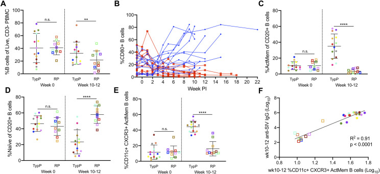
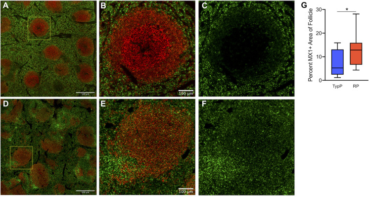
HIV-infected infants are at an increased risk of progressing rapidly to AIDS in the first weeks of life. Here, we evaluated immunological and virological parameters in 25 SIV-infected infant rhesus macaques to understand the factors influencing a rapid disease outcome. Infant macaques were infected with SIVmac251 and monitored for 10 to 17 weeks post-infection. SIV-infected infants were divided into either typical (TypP) or rapid (RP) progressor groups based on levels of plasma anti-SIV antibody and viral load, with RP infants having low SIV-specific antibodies and high viral loads. Following SIV infection, 11 out of 25 infant macaques exhibited an RP phenotype. Interestingly, TypP had lower levels of total CD4 T cells, similar reductions in CD4/CD8 ratios and elevated activation of CD8 T cells, as measured by the levels of HLA-DR, compared to RP. Differences between the two groups were identified in other immune cell populations, including a failure to expand activated memory (CD21-CD27+) B cells in peripheral blood in RP infant macaques, as well as reduced levels of germinal center (GC) B cells and T follicular helper (Tfh) cells in spleens (4- and 10-weeks post-SIV). Reduced B cell proliferation in splenic germinal GCs was associated with increased SIV+ cell density and follicular type 1 interferon (IFN)-induced immune activation. Further analyses determined that at 2-weeks post SIV infection TypP infants exhibited elevated levels of the GC-inducing chemokine CXCL13 in plasma, as well as significantly lower levels of viral envelope diversity compared to RP infants. Our findings provide evidence that early viral and immunologic events following SIV infection contributes to impairment of B cells, Tfh cells and germinal center formation, ultimately impeding the development of SIV-specific antibody responses in rapidly progressing infant macaques.


Conflict of interest statement

The authors have declared that no competing interests exist.

Figures

Fig 1
Fig 1. SIV-infected infant macaques exhibit disparate anti-SIV IgG responses.
Infants were infected with SIVmac251 either orally (n = 22) or intravenously (n = 3) with SIVmac251 and viral load and production of anti gp120 antibodies was monitored until necropsy at 10–22 weeks post infection. 11 of 25 infants that did not produce anti-gp140 specific plasma IgG or IgA during chronic SIV infection (10–12 weeks post-SIV infection) and were characterized as Rapid Progressors (RP) (A,B). RP infants failed to reduce plasma viral load after acute infection and maintained higher viral loads during chronic infection (D,E). Typical progressors (TypP) are represented as closed circles and RP infants are represented as open squares. Statistical tests used to compare infant groups were carried out as described in the methods ** = p<0.01, *** = p<0.001, **** = p<0.0001. Error bars are shown as either mean with standard deviation or median with interquartile range based on data distribution.
Fig 2
Fig 2. Number of unique plasma viral variants detected at week 2 post infection negatively correlates with anti-SIV antibodies at wk10.
SIV viral variants were assessed at 2 weeks post-infection from plasma viral RNA on an Illumina MiSeq platform. Representative phylogenies are shown for TypP (A) and RP (B) infants. The total number of unique plasma viral variants was quantified and compared between TypP (closed circles) and RP (open squares) infants (C). Number of viral variants were compared against plasma anti-SIVenv IgG concentration (D). Phylogenies were constructed using maximum likelihood with a GTR substitution model. Black lines in trees represent challenge stock consensus sequence. Statistical tests used to compare infant groups were carried out as described in the methods * = p<0.05. Error bars are shown as either mean with standard deviation or median with interquartile range based on data distribution.
Fig 3
Fig 3. Total CD4 T cell levels are sustained during chronic infection in RP infants.
CD4 T cell depletion was monitored over time using a CD4/CD8 ratio in PBMC from TypP (closed circles, blue lines) and RP (open squares, red lines) infants (A,B). The total number of peripheral CD4 T cells was monitored following SIV infection (C). Statistical tests used to compare infant groups were carried out as described in the methods * = p<0.05, ** = p<0.01, *** = p<0.001. Error bars are shown as either mean with standard deviation or median with interquartile range based on data distribution.
Fig 4
Fig 4. Activation of CD4 and CD8 T cells is increased in typically progressing infants.
Levels of HLA-DR+ CD4 (A) and CD8 (B) T cells in peripheral blood were evaluated in TypP (closed circles) and RP (open squares) infants. Both paired and unpaired and parametric and nonparametric tests were used to compare groups depending on the distribution of the data. ** = p<0.01, *** = p<0.001, **** = p<0.0001. Error bars are shown as either mean with standard deviation or median with interquartile range based on data distribution.
Fig 5
Fig 5. Rapidly progressing infants fail to increase activated memory B cells during chronic infection.
Proportions of CD20+ B cells (A) as well as CD80+ activated B cells (B) are shown from PBMC of TypP (closed circles, blue lines) and RP (open squares, red lines) infants. Proportions of activated memory (C) and naïve (D) B cell populations from total CD20+ B cells are also compared between TypP and RP infants. Within activated memory B cells we compared atypical CD11c, CXCR3 double positive cells in both TypP and RP infants (E) and correlations with anti-SIV antibody levels are shown at week 10–12 (F). Statistical tests used to compare infant groups were carried out as described in the methods ** = p<0.01, *** = p<0.001, **** = p<0.0001. Error bars are shown as either mean with standard deviation or median with interquartile range based on data distribution.
Fig 6
Fig 6. Rapidly progressing infants exhibit germinal center dysfunction in secondary lymphoid tissues.
Plasma CXCL13 concentrations were measured at timepoints prior to and following SIV infection in TypP (blue bars) and RP (red bars) infants (A) Levels of T follicular helper cells (CXCR5+, PD-1+)(B) and germinal center B cells (Ki-67+, BcL6+)(C) were measured in axillary lymph node biopsies from early chronic (4–6 weeks post-infection) and necropsy. Paraffin embedded spleens from RP and TypP infants were sectioned and immuno-stained for B cells (CD20, red) proliferation (Ki-67, green) to identify splenic B cell follicles and germinal centers. Representative images are shown for TypP infants (D) and RP infants (E). Whole sections were scanned and stitched, and Ki-67 positive foci were quantified in B cell follicles of spleen (F) and axillary lymph node (G). Statistical tests used to compare infant groups were carried out as described in the methods ** = p<0.01. Error bars are shown as either mean with standard deviation or median with interquartile range based on data distribution.
Fig 7
Fig 7. Elevated Interferon-α and pDC activation in RP infants.
Plasma IFNα concentrations were measured by ELISA at multiple time points in TypP and RP infants (A). Proportions of activated CD80+ plasmacytoid dendritic (CD123+) in PBMC from TypP (closed circles) and RP (open squares) infants were evaluated in blood (B) and lymph nodes (C) by flow cytometry. Statistical tests used to compare infant groups were carried out as described in the methods * = p<0.05, *** = p<0.001, **** = p<0.0001. Error bars are shown as either mean with standard deviation or median with interquartile range based on data distribution.
Fig 8
Fig 8. Increased type 1 IFN associated protein expression is observed in B cell follicles of RP infants.
Levels of MX1 protein were measured in areas within B cell follicles of spleen. Paraffin embedded spleens from RP and TypP infants were sectioned and immuno-stained for B cells (CD20, red) and MX1 (green) to identify splenic B cell follicles and IFN-induced protein expression. Representative images are shown for TypP infants (A-C) and RP infants (D-F). Whole sections were scanned and stitched and MX1 was quantified within 10 randomly selected splenic B cell follicles (G) for TypP (blue bar) and RP (red bar) infants. Statistical tests used to compare infant groups were carried out as described in the methods * = p<0.05.
Fig 9
Fig 9. Rapidly progressing infants have more SIV infected cells localized outside of splenic germinal centers.
RNAscope in situ hybridization using SIV-specific RNA probes was used to detect SIV-infected cells and cell-free virus in the spleens of TypP (A) and RP (B) infants. SIV+ cells were quantified across stitched images and normalized for area of splenic tissue (C). Statistical tests used to compare infant groups were carried out as described in the methods * = p<0.05. Error bars are shown as floating bars (min to max) with line indicating the mean. n = 3 per group.
Fig 10
Fig 10. Model summarizing factors influencing rapid SIV progression in infants
Illustration of events following infection in infant macaques exhibiting rapid disease progression. GC = germinal center; Tfh = T follicular helper cell; pDC = plasmacytoid dendritic cell.

Similar articles

Cited by

References

    1. UNAIDS. AIDSinfo 2019 [cited 2019. Available from: aidsinfo.unaids.org.
    1. Newell ML, Coovadia H, Cortina-Borja M, Rollins N, Gaillard P, Dabis F, et al.. Mortality of infected and uninfected infants born to HIV-infected mothers in Africa: a pooled analysis. Lancet. 2004;364(9441):1236–43, 10.1016/S0140-6736(04)17140-7, https://www.ncbi.nlm.nih.gov/pubmed/15464184. - DOI - PubMed
    1. Espanol T, Garcia-Armui R, Bofill A, Sune J, Bertran JM. Hypogammaglobulinaemia and negative anti-HIV antibodies in AIDS. Arch Dis Child. 1987;62(8):853–4, 10.1136/adc.62.8.853, https://www.ncbi.nlm.nih.gov/pubmed/3662596. - DOI - PMC - PubMed
    1. Pahwa R, Good RA, Pahwa S. Prematurity, hypogammaglobulinemia, and neuropathology with human immunodeficiency virus (HIV) infection. Proc Natl Acad Sci U S A. 1987;84(11):3826–30, 10.1073/pnas.84.11.3826, https://www.ncbi.nlm.nih.gov/pubmed/3473485. - DOI - PMC - PubMed
    1. Gaetano C, Scano G, Carbonari M, Giannini G, Mezzaroma I, Aiuti F, et al.. Delayed and defective anti-HIV IgM response in infants. Lancet. 1987;1(8533):631, 10.1016/s0140-6736(87)90272-8, https://www.ncbi.nlm.nih.gov/pubmed/2881166. - DOI - PubMed

Publication types

MeSH terms

Substances

LinkOut - more resources