. 2021 Jul:155:105388.
doi: 10.1016/j.nbd.2021.105388. Epub 2021 May 5.

Extracellular vesicles regulate gap junction-mediated intercellular communication and HIV-1 infection of human neural progenitor cells

Affiliations

Extracellular vesicles regulate gap junction-mediated intercellular communication and HIV-1 infection of human neural progenitor cells

Hyung Joon Cho et al. Neurobiol Dis. 2021 Jul.

Abstract

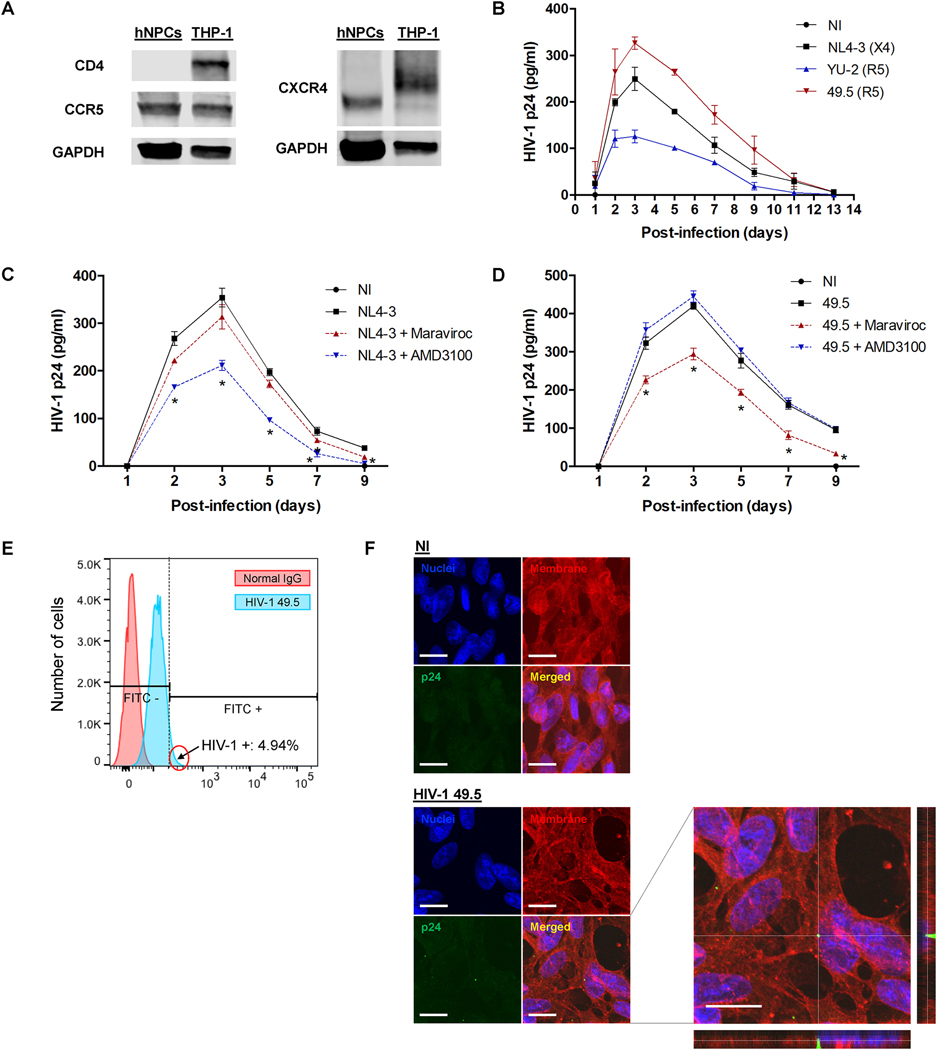
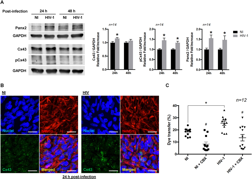
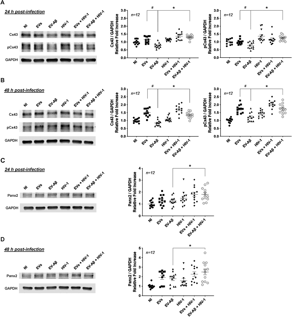
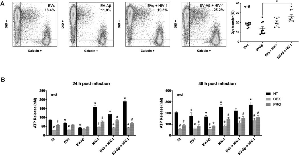
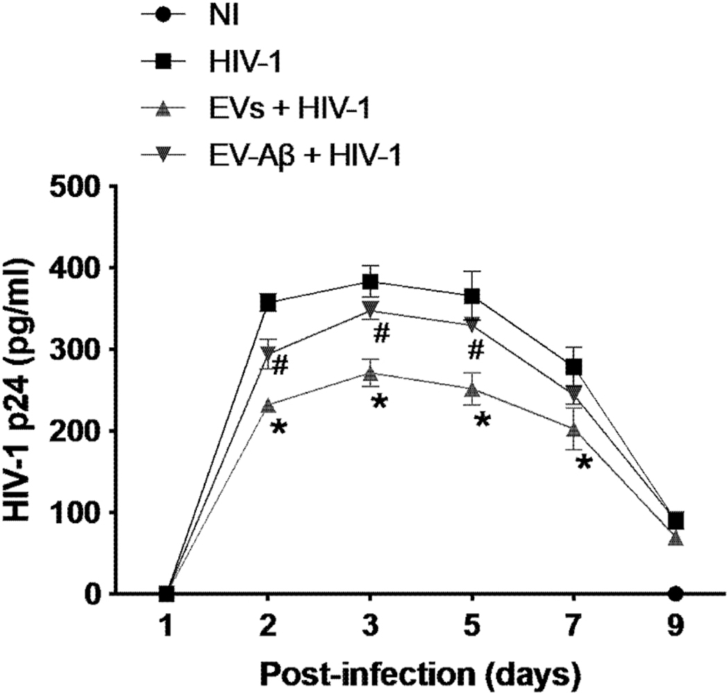
Human immunodeficiency virus-1 (HIV-1) has been shown to cross the blood-brain barrier and cause HIV-associated neurocognitive disorders (HAND) through a process that may involve direct or indirect interactions with the central nervous system (CNS) cells and alterations of amyloid β (Aβ) homeostasis. The present study focused on the mechanisms of HIV-1 infecting human neural progenitor cells (hNPCs) and affecting NPC intercellular communications with human brain endothelial cells (HBMEC). Despite the lack of the CD4 receptor, hNPCs were effectively infected by HIV-1 via a mechanism involving the chemokine receptors, CXCR4 and CCR5. HIV-1 infection increased expression of connexin-43 (Cx43), phosphorylated Cx43 (pCx43), and pannexin 2 (Panx2) protein levels in hNPCs, suggesting alterations in gap-junction (GJ) and pannexin channel communication. Indeed, a functional GJ assay indicated an increase in communication between HIV-infected hNPCs and non-infected HBMEC. We next analyzed the impact of HBMEC-derived extracellular vesicles (EVs) and EVs carrying Aβ (EV-Aβ) on the expression of Cx43, pCx43, and Panx2 in HIV-1 infected and non-infected hNPCs. Exposure to EV-Aβ resulted in significant reduction of Cx43 and pCx43 protein expression in non-infected hNPCs when compared to EV controls. Interestingly, EV-Aβ treatment significantly increased levels of Cx43, pCx43, and Panx2 in HIV-1-infected hNPCs when compared to non-infected controls. These results were confirmed in a GJ functional assay and an ATP release assay, which is an indicator of connexin hemichannel and/or pannexin channel functions. Overall, the current study demonstrates the importance of hNPCs in HIV-1 infection and indicates that intercellular communications between infected hNPCs and HBMEC can be effectively modulated by EVs carrying Aβ as their cargo.

Keywords: Amyloid β; Connexins; Extracellular vesicles; HIV-1; Human neural progenitor cells; Pannexins.


Conflict of interest statement

Declaration of Competing Interest

The authors declare no conflict of interest.

Figures

Figure 1.
Figure 1.. Human neural progenitor cells (hNPCs) can be infected by both the X4 and R5 tropic HIV-1 strains.
(A) Protein expression of HIV-1 receptors CD4, CXCR4 and CCR5 in untreated hNPCs. hNPCs express the CXCR4 and CCR5 HIV-1 co-receptors but not the CD4 receptor. A human monocytic leukemia cell line (THP-1) was used as a positive control. (B) Quantification of p24 release into NPC culture media following infection with the X4 and R5 tropic HIV-1 strains. Cells were infected with HIV-1 NL4–3 (X4), HIV-1 YU-2, or 49.5 (both R5) and media was collected at the indicated times. Data represents mean ± SEM (n=4) from three independent experiments (total n=12 per group). (C and D) Impact of CXCR4 or CCR5 inhibition on HIV-1 infection in hNPCs. hNPCs were pretreated with CXCR4 antagonist AMD3100 or CCR5 antagonist Maraviroc (both at 1μM for 3 h) and then infected with (C) NL4–3 (X4) or (D) 49.5 (R5) HIV-1 strain in the presence of AMD3100 or CCR5. p24 levels in the media were assessed as a marker of active HIV-1 replication. Note that AMD3100 effectively protected against NPC infection by the NL4–3 strain and Maraviroc against infection by the 49.5 strain. Data represents mean ± SEM for a representative experiment (n=4) from three independent experiments (total n=12 per group). (E) Quantification of HIV-1 infectivity in hNPCs. hNPCs were infected with HIV-1 49.5 for 24 h; then, the population of HIV-1 p24 positive hNPCs was quantitatively measured by flow cytometry. The results indicate about 5% of HIV-1-infected hNPCs. The histogram is a representative flow cytometry result of n=4. (F) Representative images of p24 immunoreactivity. NPCs were infected with HIV-1 49.5 strain for 48 h, followed by staining for p24 (green) as a marker of productive HIV-1 infection, for cell membrane (red, DiI staining) and nuclei (blue, DAPI staining. No p24 levels were detected in non-infected (NI) group. Scale bar: 20 μm.
Figure 2.
Figure 2.. HIV-1 infection upregulates gap junction (GJ) and hemichannel proteins.
(A) Connexin 43 (Cx43), phosphorylated Cx43 (pCx43), and pannexin 2 (Panx2) protein levels in hNPCs infected with the 49.5 HIV strain for 24 or 48 h. Left panel shows representative Western blots and right panel reflects quantitative results of these analyses as compared to the non-infected (NI) group (n=14 from four independent experiments). All target proteins (Cx43, pCx43, and Panx2) were normalized to a reference target (GAPDH). (B) Representative images of Cx43 distribution in HIV-1-infected hNPCs. hNPC were infected with HIV-1 49.5 for 24 h. Non-infected cells were used as control. Note stronger Cx43 immunoreactivity in HIV-1-infected cultures. Scale bar: 20 μm. (C) Gap junction (GJ) channel function between HIV-1-infected hNPCs and non-infected HBMECs. hNPCs were infected with HIV-1 (the 49.5 strain) for 24 h in the presence or absence of carbenoxolone (CBX; 100 μM), and GJ-mediated dye-coupling transfer towards non-infected HBMECs was assessed by flow cytometry. Non-infected (NI) cells served as control. Donor hNPCs were labeled with calcein and recipient HBMECs with DiD. Quantitative analysis of the flow cytometry results demonstrated that HIV infection opens GJ channel function. Scatter dot plots show individual values with mean ± SEM (n=12 from four independent experiments). *p<0.05 vs. NI. #p < 0.05 vs. no CBX in NI or HIV-1-infected cells.
Figure 3.
Figure 3.. Impact of extracellular vesicles (EVs) and EV-associated Aβ (EV-Aβ) on GJ and hemichannel proteins in HIV-1-infected hNPCs.
hNPCs were pretreated with EVs or EV-Aβ for 3 h and infected with HIV-1 49.5 in the presence of EVs or EV-Aβ for 24 h (A and C) and 48 h (B and D). Protein expression of Cx43, pCx43 (A and B), and Panx2 (C and D) were analyzed by immunoblotting. Left panels show representative immunoblots and right panels provides quantitative results of these analyses as compared to the control group. Scatter dot plots show individual values with mean ± SEM (n=12 from three independent experiments). All target proteins (Cx43, pCx43, and Panx2) were normalized to a reference target (GAPDH). NI: non-infected cells.
Figure 4.
Figure 4.. Impact of EVs and EV-Aβ on GJ and hemichannel function in HIV-1-infected hNPCs.
(A) GJ channel function. hNPC were treated with EVs or EV-Aβ and infected with HIV-1 as in Figure 3, followed by the analysis of dye transfer to non-infected HBMEC as in Figure 2C. Left panel, a representative flow cytometry measurement. Right panel, scatter dot plots show individual values with mean ± SEM (n = 9 from three independent experiments). *p < 0.05 vs. EV-Aβ. (B) Hemichannel activity. hNPC were treated as in (A). In addition, cells were exposed to carbenoxolone (CBX, a connexin blocker, 25 μM) or probenecid (PRO, a pannexin hemichannel blocker, 0.5 mM). Hemichannel activity was measured by the assessment of ATP release into cell culture media after 24 h (left panel) and 48 h (right panel). NI: non-infected cells; NT: no treatment. Data represents mean ± SEM (n=8 from three independent experiments). *p < 0.05 vs. NI; #p < 0.05 vs. NT of each group.
Figure 5.
Figure 5.. Kinetics of HIV-1 infection in EVs or EV-Aβ pretreated hNPCs.
Quantification of p24 levels in the culture media were assessed as in Figure 1 in the presence of EVs or EV-Aβ. Data represents mean ± SEM for a representative experiment (n=4) from three independent experiments (total n=12 per group). *p < 0.05 vs. HIV-1 and #p < 0.05 vs. EVs + HIV-1.

Similar articles

Cited by

References

    1. Achim CL, Adame A, Dumaop W, Everall IP, Masliah E, Neurobehavioral Research C, 2009. Increased accumulation of intraneuronal amyloid β in HIV-infected patients. J Neuroimmune Pharmacol. 4, 190–199. - PMC - PubMed
    1. Alvarez Losada S, Cantó-Nogués C, Muñoz-Fernández MA, 2002. A new possible mechanism of human immunodeficiency virus type 1 infection of neural cells. Neurobiol Dis. 11, 469–478. - PubMed
    1. András IE, Garcia-Contreras M, Yanick C, Perez P, Sewell B, Durand L, Toborek M, 2020a. Extracellular vesicle-mediated amyloid transfer to neural progenitor cells: implications for RAGE and HIV infection. Mol Brain. 13, 21. - PMC - PubMed
    1. András IE, Leda A, Contreras MG, Bertrand L, Park M, Skowronska M, Toborek M, 2017. Extracellular vesicles of the blood-brain barrier: Role in the HIV-1 associated amyloid beta pathology. Mol Cell Neurosci. 79, 12–22. - PMC - PubMed
    1. András IE, Sewell BB, Toborek M, 2020b. HIV-1 and Amyloid Beta Remodel Proteome of Brain Endothelial Extracellular Vesicles. Int J Mol Sci. 21. - PMC - PubMed

Publication types

MeSH terms

Substances