Review
. 2021 Oct:52:101245.
doi: 10.1016/j.molmet.2021.101245. Epub 2021 May 4.

Insulin-like growth factors: Ligands, binding proteins, and receptors

Affiliations
Review

Insulin-like growth factors: Ligands, binding proteins, and receptors

Derek LeRoith et al. Mol Metab. 2021 Oct.

Abstract

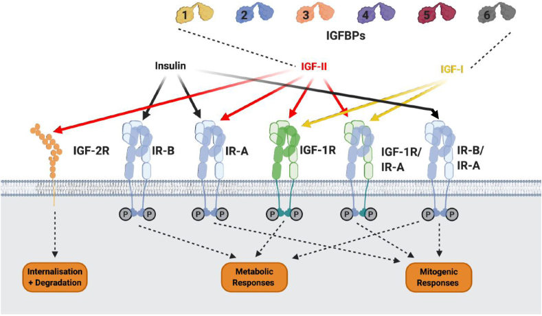
Background: The insulin-like growth factor family of ligands (IGF-I, IGF-II, and insulin), receptors (IGF-IR, M6P/IGF-IIR, and insulin receptor [IR]), and IGF-binding proteins (IGFBP-1-6) play critical roles in normal human physiology and disease states.

Scope of review: Insulin and insulin receptors are the focus of other chapters in this series and will therefore not be discussed further. Here we review the basic components of the IGF system, their role in normal physiology and in critical pathology's. While this review concentrates on the role of IGFs in human physiology, animal models have been essential in providing understanding of the IGF system, and its regulation, and are briefly described.

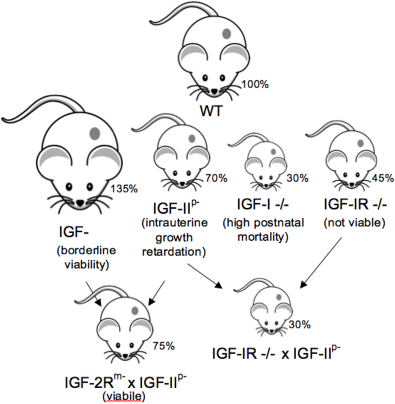
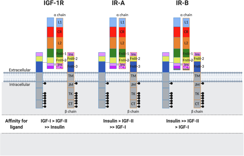
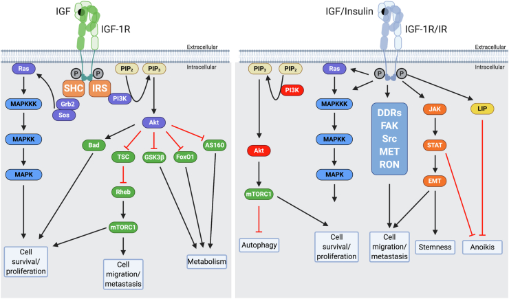
Major conclusions: IGF-I has effects via the circulation and locally within tissues to regulate cellular growth, differentiation, and survival, thereby controlling overall body growth. IGF-II levels are highest prenatally when it has important effects on growth. In adults, IGF-II plays important tissue-specific roles, including the maintenance of stem cell populations. Although the IGF-IR is closely related to the IR it has distinct physiological roles both on the cell surface and in the nucleus. The M6P/IGF-IIR, in contrast, is distinct and acts as a scavenger by mediating internalization and degradation of IGF-II. The IGFBPs bind IGF-I and IGF-II in the circulation to prolong their half-lives and modulate tissue access, thereby controlling IGF function. IGFBPs also have IGF ligand-independent cell effects.

Keywords: Cancer; Growth; Insulin-like growth factor binding proteins; Insulin-like growth factor receptors; Insulin-like growth factors; Metabolism.


Figures

Figure 1
Figure 1
Summary of mouse studies investigating IGF system genes and their role in somatic growth. Percentages indicate body weight relative to age-matched normal mice. M indicates maternally disrupted allele, P indicates paternally disrupted allele and −/− indicates both alleles disrupted.
Figure 2
Figure 2
Major components of the IGF system. The three insulin peptides, IGF-I, and IGF-II act via a series of cell surface receptors. The availability of IGF-I and IGF-II but not insulin to bind to these receptors is modulated via binding to six soluble high-affinity binding proteins, IGFBP-1 to −6. IGF-2R also modulates the availability of IGF-II by binding, internalizing, and targeting IGF-II for degradation. Insulin binds with the highest affinity to the two insulin receptor isoforms IR-A and IR-B. IR-A and IR-B are splice variants of the IR, with IR-A having a modified ligand-binding domain due to exon 11 being spliced out. Insulin has a low affinity for the IGF-IR. Each “hemireceptor” of IR-A, IR-B, and IGF-IR are comprised of an α- and β-subunit; each of these hemireceptors can either homodimerize or heterodimerize, forming hybrid receptors. IGF-I binds with a high affinity to the IGF-IR, IGF-IR/IR-A, and IGF-IR/IR-B hybrid receptors. IGF-II binds with high affinity to the IGF-IR, IR-A, and IGF-IR/IR-A hybrid receptors. Insulin only binds to hybrid receptors with low affinity. Activation of IR-B, IGF-1R, and IR-A/IR-B hybrids elicits metabolic responses. Activation of IR-A, IGF-IR, IGF-IR/IR-A, and IR-A/IR-B hybrids elicits mitogenic responses.
Figure 3
Figure 3
The IGF-1R and IR structural domains and affinities. L1 and L2: leucine-rich repeat domains; CR: cysteine-rich region; FnIII-1, FnIII-2, and FnIII-3: fibronectin-type III domains; Ins: insertion domains (α-chain C-terminal component and β-chain component); TM: transmembrane region; JM: juxtamembrane region; TK: tyrosine kinase catalytic domain; CT: C-terminal region.
Figure 4
Figure 4
Left panel: Canonical IGF-I and IGF-II signaling through the IGF-IR. IGF-I and IGF-II bind the IGF-IR at the extracellular α subunit, leading to autophosphorylation of β subunit residues, which then act as docking sites for insulin receptor substrates (IRS) 1–4 and other signaling proteins, such Shc. IRS-1 recruits the p85 regulatory subunit of phosphatidylinositol 3-kinase (PI3K), which through the catalytic effects of its p110 subunit recruits protein kinase B (Akt) to the cell membrane, where it is phosphorylated and activated. Activated Akt has many substrates, including Bcl2 antagonist of cell death (Bad), glycogen synthase kinase 3β (GSK3β), forkhead transcription factors (such as FoxO1), and Akt substrate of 160 kDa (AS160). These factors are chiefly involved in regulating apoptosis and cell metabolism. Akt also regulates protein synthesis by phosphorylating tuberous sclerosis protein (TSC2), releasing its inhibition of Rheb to activate the mammalian target of rapamycin (mTORC1). The phosphorylation of IRS-1 and Shc can also lead to recruitment of growth factor receptor-bound protein 2 (Grb2), Son of Sevenless (SOS), and Ras, with subsequent activation via a phosphorylation series of the mitogen-activated protein kinase (MAPKKK, MAPKK, and MAPK) pathways, leading to cell proliferation, migration, and survival. Right panel: More novel signaling through the IGF-IR and IR mediated via additional interactions of the C-terminal intracellular region of the activated receptors with factors such as discoidin domain receptors (DDRs), focal adhesion kinase (FAK), receptor tyrosine-protein kinases such as Src, mesenchymal epithelial transition factor (MET), and recepteur d'origine nantais (RON). The activated receptor can also interact with Janus kinase (JAK), signal transducer, and activator of transcription (STAT). These interactions contribute to receptor actions on autophagy, epithelial–mesenchymal transition (EMT), stemness, and anoikis.

References

    1. Salmon W.D., Jr., Daughaday W.H. A hormonally controlled serum factor which stimulates sulfate incorporation by cartilage in vitro. The Journal of Laboratory and Clinical Medicine. 1957;49(6):825–836. - PubMed
    1. Argon Y., Bresson S.E., Marzec M.T., Grimberg A. Glucose-regulated protein 94 (GRP94): a novel regulator of insulin-like growth factor production. Cells. 2020;9(8):1844. doi: 10.3390/cells9081844. - DOI - PMC - PubMed
    1. Roed N.K., Viola C.M., Kristensen O., Schluckebier G., Norrman M., Sajid W. Structures of insect Imp-L2 suggest an alternative strategy for regulating the bioavailability of insulin-like hormones. Nature Communications. 2018;9(1):3860. doi: 10.1038/s41467-018-06192-3. - DOI - PMC - PubMed
    1. Yakar S., Isaksson O. Regulation of skeletal growth and mineral acquisition by the GH/IGF-1 axis: lessons from mouse models. Growth Hormone & IGF Research. 2016;28:26–42. - PMC - PubMed
    1. Powell-Braxton L., Hollingshead P., Warburton C., Dowd M., Pitts-Meek S., Dalton D. IGF-I is required for normal embryonic growth in mice. Genes & Development. 1993;7(12B):2609–2617. - PubMed