

Response patterns in adventitial layer of Echinococcus granulosus sensu stricto cysts from naturally infected cattle and sheep
- PMID: 33962666
- PMCID: PMC8105927
- DOI: 10.1186/s13567-021-00936-8
Response patterns in adventitial layer of Echinococcus granulosus sensu stricto cysts from naturally infected cattle and sheep
Abstract
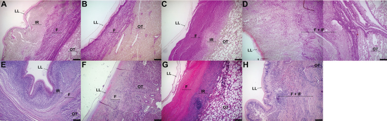
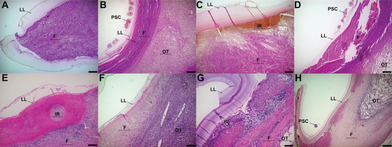
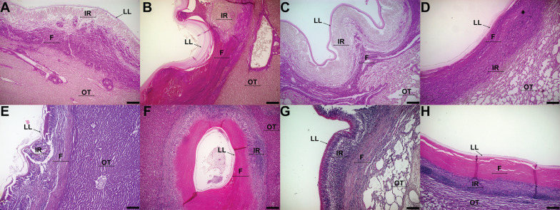
Cystic echinococcosis is a zoonotic disease caused by the metacestode of Echinococcus granulosus sensu lato. The disease is characterized by the development of cystic structures inside viscera of the intermediate host, mainly liver and lungs. These cysts are formed by three layers: germinal, laminated, and adventitial layer, the latter being the local host immune response. Metacestodes that develop protoscoleces, the infective stage to the definitive host, are termed fertile, whereas cysts that do not produce protoscoleces are termed non-fertile. Sheep usually harbor fertile cysts while cattle usually harbor non-fertile cysts. Adventitial layers with fibrotic resolution are associated to fertile cysts, whereas a granulomatous reaction is associated with non-fertile cysts. The aim of this study was to analyze cellular distribution in the adventitial layer of fertile and non-fertile E. granulosus sensu stricto cysts found in liver and lungs of cattle and sheep. A total of 418 cysts were analyzed, 203 from cattle (8 fertile and 195 non-fertile) and 215 from sheep (64 fertile and 151 non-fertile). Fertile cysts from cattle showed mixed patterns of response, with fibrotic resolution and presence of granulomatous response in direct contact with the laminated layer, while sheep fertile cysts always displayed fibrotic resolution next to the laminated layer. Cattle non-fertile cysts display a granulomatous reaction in direct contact with the laminated layer, whereas sheep non-fertile cysts display a granulomatous reaction, but in direct contact with the fibrotic resolution. This shows that cattle and sheep cystic echinococcosis cysts have distinct local immune response patterns, which are associated to metacestode fertility.
Keywords: Adventitial layer; Echinococcus granulosus; Fibrosis; Histopathology.
Conflict of interest statement
The authors declare that they have no competing interests.
Figures
Similar articles
-
Transcriptome analysis of Echinococcus granulosus sensu stricto protoscoleces reveals differences in immune modulation gene expression between cysts found in cattle and sheep.Vet Res. 2022 Jan 28;53(1):8. doi: 10.1186/s13567-022-01022-3. Vet Res. 2022. PMID: 35090558 Free PMC article.
-
Fasciola hepatica coinfection modifies the morphological and immunological features of Echinococcus granulosus cysts in cattle.Vet Res. 2020 Jun 5;51(1):76. doi: 10.1186/s13567-020-00799-5. Vet Res. 2020. PMID: 32503674 Free PMC article.
-
Multiple haplotypes of Echinococcus granulosus sensu stricto in single naturally infected intermediate hosts.Parasitol Res. 2020 Feb;119(2):763-770. doi: 10.1007/s00436-019-06578-2. Epub 2019 Dec 20. Parasitol Res. 2020. PMID: 31863181
-
Echinococcus granulosus sensu lato in animal intermediate hosts: What is with the organ location?Vet Parasitol. 2022 Apr;304:109695. doi: 10.1016/j.vetpar.2022.109695. Epub 2022 Mar 9. Vet Parasitol. 2022. PMID: 35298991 Review.
-
Hepatic echinococcosis: clinical and therapeutic aspects.World J Gastroenterol. 2012 Apr 7;18(13):1448-58. doi: 10.3748/wjg.v18.i13.1448. World J Gastroenterol. 2012. PMID: 22509076 Free PMC article. Review.
Cited by
-
HLA class II profile in patients with different stages of cystic echinococcosis according to the WHO ultrasound imaging classification.Parasitol Res. 2024 Sep 24;123(9):330. doi: 10.1007/s00436-024-08353-4. Parasitol Res. 2024. PMID: 39316156
-
Zoonotic threat of cystic echinococcosis in Tunisia: insights into livestock prevalence and identification of the G1 genotype.Front Vet Sci. 2025 Feb 20;12:1536368. doi: 10.3389/fvets.2025.1536368. eCollection 2025. Front Vet Sci. 2025. PMID: 40051979 Free PMC article.
-
Weighted gene co-expression network analysis reveals immune evasion related genes in Echinococcus granulosus sensu stricto.Exp Biol Med (Maywood). 2024 Feb 29;249:10126. doi: 10.3389/ebm.2024.10126. eCollection 2024. Exp Biol Med (Maywood). 2024. PMID: 38510493 Free PMC article.
-
Transcriptome analysis of Echinococcus granulosus sensu stricto protoscoleces reveals differences in immune modulation gene expression between cysts found in cattle and sheep.Vet Res. 2022 Jan 28;53(1):8. doi: 10.1186/s13567-022-01022-3. Vet Res. 2022. PMID: 35090558 Free PMC article.
-
Metabolomics Analysis of Splenic CD19+ B Cells in Mice Chronically Infected With Echinococcus granulosus sensu lato Protoscoleces.Front Vet Sci. 2021 Sep 6;8:718743. doi: 10.3389/fvets.2021.718743. eCollection 2021. Front Vet Sci. 2021. PMID: 34552973 Free PMC article.
References
MeSH terms
Grants and funding
LinkOut - more resources
Full Text Sources
Other Literature Sources
