

Premedication with pioglitazone prevents doxorubicin-induced left ventricular dysfunction in mice
- PMID: 33962676
- PMCID: PMC8103594
- DOI: 10.1186/s40360-021-00495-w
Premedication with pioglitazone prevents doxorubicin-induced left ventricular dysfunction in mice
Abstract
Background: Doxorubicin (DOX) is widely used as an effective chemotherapeutic agent for cancers; however, DOX induces cardiac toxicity, called DOX-induced cardiomyopathy. Although DOX-induced cardiomyopathy is known to be associated with a high cumulative dose of DOX, the mechanisms of its long-term effects have not been completely elucidated. Pioglitazone (Pio) is presently contraindicated in patients with symptomatic heart failure owing to the side effects. The concept of drug repositioning led us to hypothesize the potential effects of Pio as a premedication before DOX treatment, and to analyze this hypothesis in mice.
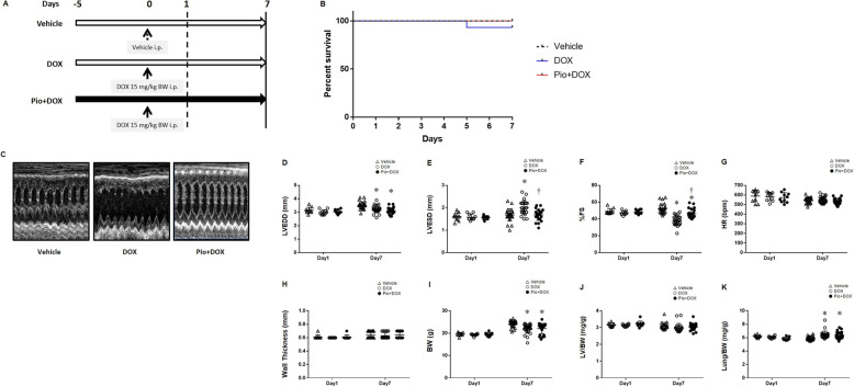
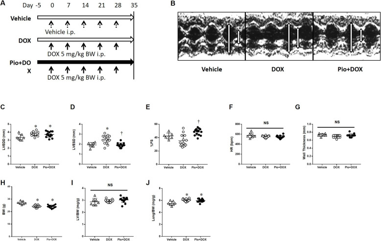
Methods: First, for the hyperacute (day 1) and acute (day 7) DOX-induced dysfunction models, mice were fed a standard diet with or without 0.02% (wt/wt) Pio for 5 days before DOX treatment (15 mg/kg body weight [BW] via intraperitoneal [i.p.] administration). The following 3 treatment groups were analyzed: standard diet + vehicle (Vehicle), standard diet + DOX (DOX), and Pio + DOX. Next, for the chronic model (day 35), the mice were administrated DOX once a week for 5 weeks (5 mg/kg BW/week, i.p.).
Results: In the acute phase after DOX treatment, the percent fractional shortening of the left ventricle (LV) was significantly decreased in DOX mice. This cardiac malfunction was improved in Pio + DOX mice. In the chronic phase, we observed that LV function was preserved in Pio + DOX mice.
Conclusions: Our findings may provide a new pathophysiological explanation by which Pio plays a role in the treatment of DOX-induced cardiomyopathy, but the molecular links between Pio and DOX-induced LV dysfunction remain largely elusive.
Keywords: Anticancer agent; Cardiac toxicity; LV dysfunction; Thiazolidinediones.
Conflict of interest statement
The authors declare that they have no conflicts of interest associated with this manuscript.
Figures
Similar articles
-
Long-acting PDE5 inhibitor tadalafil prevents early doxorubicin-induced left ventricle diastolic dysfunction in juvenile mice: potential role of cytoskeletal proteins.Can J Physiol Pharmacol. 2017 Mar;95(3):295-304. doi: 10.1139/cjpp-2016-0551. Epub 2017 Feb 26. Can J Physiol Pharmacol. 2017. PMID: 28238269
-
Granulocyte colony-stimulating factor improves left ventricular function of doxorubicin-induced cardiomyopathy.Lab Invest. 2007 May;87(5):440-55. doi: 10.1038/labinvest.3700530. Epub 2007 Mar 5. Lab Invest. 2007. PMID: 17334414
-
Pioglitazone modulates doxorubicin resistance in a in vivo model of drug resistant osteosarcoma xenograft.Naunyn Schmiedebergs Arch Pharmacol. 2021 Feb;394(2):361-371. doi: 10.1007/s00210-020-01982-3. Epub 2020 Oct 5. Naunyn Schmiedebergs Arch Pharmacol. 2021. PMID: 33015747
-
Dietary inorganic nitrate alleviates doxorubicin cardiotoxicity: mechanisms and implications.Nitric Oxide. 2012 May 15;26(4):274-84. doi: 10.1016/j.niox.2012.03.006. Epub 2012 Apr 5. Nitric Oxide. 2012. PMID: 22484629 Free PMC article. Review.
-
A concise description of cardioprotective strategies in doxorubicin-induced cardiotoxicity.Can J Physiol Pharmacol. 2009 Oct;87(10):756-63. doi: 10.1139/Y09-059. Can J Physiol Pharmacol. 2009. PMID: 19898559 Review.
Cited by
-
Cardioprotection strategies for anthracycline cardiotoxicity.Basic Res Cardiol. 2025 Feb;120(1):71-90. doi: 10.1007/s00395-024-01078-6. Epub 2024 Sep 9. Basic Res Cardiol. 2025. PMID: 39249555 Free PMC article. Review.
-
Anti-Diabetic Therapies and Cancer: From Bench to Bedside.Biomolecules. 2024 Nov 20;14(11):1479. doi: 10.3390/biom14111479. Biomolecules. 2024. PMID: 39595655 Free PMC article. Review.
-
Cross-disease communication between cancer and heart failure provides a rational approach to prevention and treatment of both diseases.Front Oncol. 2022 Oct 31;12:1006322. doi: 10.3389/fonc.2022.1006322. eCollection 2022. Front Oncol. 2022. PMID: 36387253 Free PMC article. Review.
References
-
- Cardinale D, Colombo A, Lamantia G, Colombo N, Civelli M, De Giacomi G, Rubino M, Veglia F, Fiorentini C, Cipolla CM. Anthracycline-induced cardiomyopathy: clinical relevance and response to pharmacologic therapy. J Am Coll Cardiol. 2010;55(3):213–220. doi: 10.1016/j.jacc.2009.03.095. - DOI - PubMed
-
- Zamorano JL, Lancellotti P, Rodriguez Munoz D, Aboyans V, Asteggiano R, Galderisi M, Habib G, Lenihan DJ, Lip GYH, Lyon AR, et al. 2016 ESC position paper on cancer treatments and cardiovascular toxicity developed under the auspices of the ESC Committee for practice guidelines: the task force for cancer treatments and cardiovascular toxicity of the European Society of Cardiology (ESC) Eur Heart J. 2016;37(36):2768–2801. doi: 10.1093/eurheartj/ehw211. - DOI - PubMed
Publication types
MeSH terms
Substances
LinkOut - more resources
Full Text Sources
Other Literature Sources
