Review
. 2021:360:65-98.
doi: 10.1016/bs.ircmb.2020.09.004. Epub 2020 Nov 24.

Actin dynamics during tumor cell dissemination

Affiliations
Review

Actin dynamics during tumor cell dissemination

Chandrani Mondal et al. Int Rev Cell Mol Biol. 2021.

Abstract

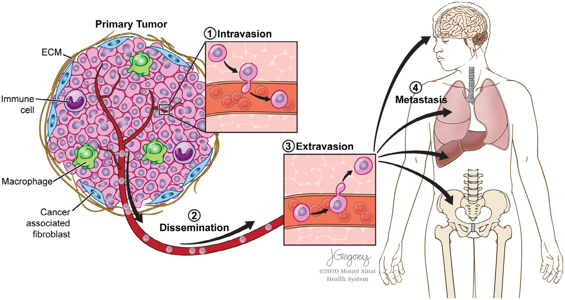
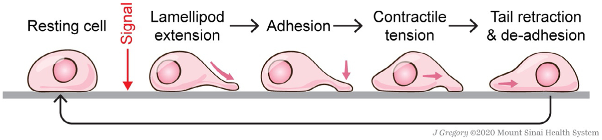
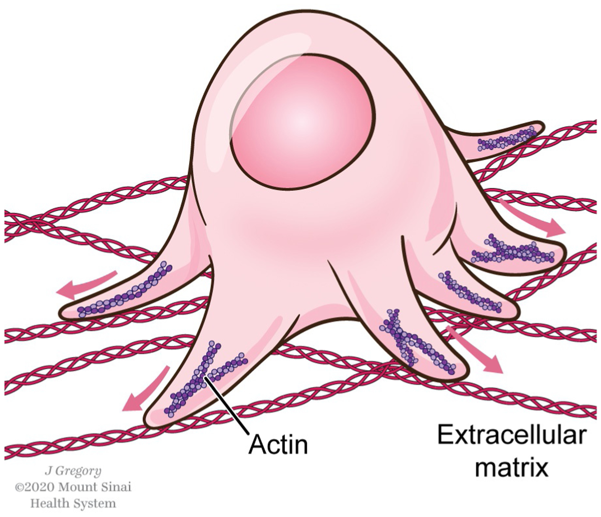
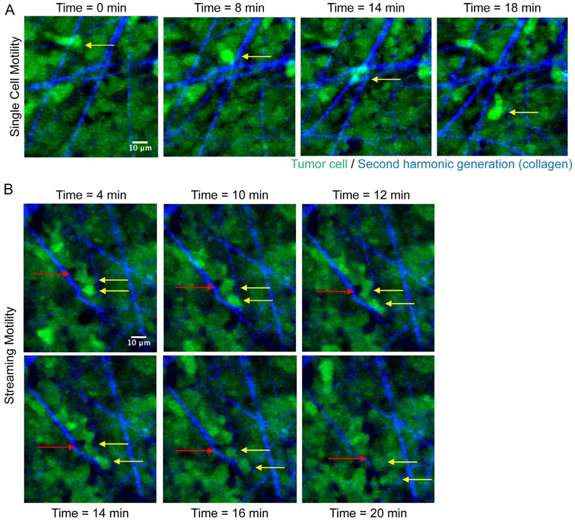
The actin cytoskeleton is a dynamic network that regulates cellular behavior from development to disease. By rearranging the actin cytoskeleton, cells are capable of migrating and invading during developmental processes; however, many of these cellular properties are hijacked by cancer cells to escape primary tumors and disseminate to distant organs in the body. In this review article, we highlight recent work describing how cancer cells regulate the actin cytoskeleton to achieve efficient invasion and metastatic colonization. We also review new imaging technologies that are capable of revealing the complex architecture and regulation of the actin cytoskeleton during motility and invasion of tumor cells.

Keywords: Actin cytoskeleton; Cancer cell motility and dissemination; Tumor microenvironment.


Figures

Fig. 1
Fig. 1
Invasion-metastasis cascade. Primary tumors have a complex tumor microenvironment (e.g., immune cells, cancer-associated fibroblasts, the extracellular matrix) that plays a dynamic role in affecting tumor cell dissemination and how tumor cells intravasate into the vasculature (1), disseminate through the circulatory system (2), extravasate out of the vasculature (3), and colonize distant organs (4).
Fig. 2
Fig. 2
Cell motility cycle. Upon integrating signaling cues, a resting cell can form lamellipodia, or protrusive actin-rich structures, and adhere to the substrata with nascent adhesions. In order for the cell body to translocate, the cell experiences contractile tension and rear detachment through de-adhesion.
Fig. 3
Fig. 3
Tumor cell in a 3D context. In the tumor microenvironment, cancer cells encounter a complex extracellular matrix, and are capable of forming different types of protrusive structures, including pseudopodia.
Fig. 4
Fig. 4
Intravital imaging of a melanoma xenograft. Two-photon imaging of tumor cells (in green) and second harmonic generation of fibrillar collagen (in blue). (A) Yellow arrow points to a single cell moving over time (indicated above each panel). (B) Example of streaming motility; yellow arrows point to cancer cells following each other, and red arrow points to another cell type in the tumor microenvironment moving within the multicellular stream. Scale Bar: 10μm. SK-Mel-147 GFP-labeled melanoma cells were injected subcutaneously in 6-week old female nude mice and tumors were allowed to grow up to 1cm3. Intravital imaging of the primary tumor was performed as in Patsialou et al. (2013) and collagen fibers were visualized by second harmonic generation. Images were acquired every 2min for 30min, with a step-size of 5μm.
Fig. 5
Fig. 5
dSTORM image of a breast cancer cell. (Left panel): Widefield image of a cancer cell on gelatin matrix. F-actin in blue, cortactin in red. (Middle panel): dSTORM image of the same cancer cell. Top and bottom inset #1 (yellow box): Widefield and dSTORM of a lamellipodia structure. Top and bottom inset #2 (green box): Widefield and dSTORM showing spatial distribution of F-actin and cortactin in an invadopodia structure. Left and middle panel, scale bar: 10μm. Insets, scale bar: 1μm. Data acquisition for dSTORM was carried out on the Nanoimager S (Oxford NanoImaging, ONI, Oxford, UK). Signals from Alexa 647 and Alexa 488 were recorded sequentially for 10,000 frames each. Localization and image rendering were performed in the NimOS v1.4 software, and the final reconstruction displayed in a precision mode.

References

    1. Aiello NM, Kang Y, 2019. Context-dependent EMT programs in cancer metastasis. J. Exp. Med 216 (5), 1016–1026. 10.1084/jem.20181827. Rockefeller University Press. - DOI - PMC - PubMed
    1. Alieva M, et al., 2019. Intravital imaging of glioma border morphology reveals distinctive cellular dynamics and contribution to tumor cell invasion. Sci. Rep 9 (1), 2054. 10.1038/s41598-019-38625-4. Nature Publishing Group UK. - DOI - PMC - PubMed
    1. Arjonen A, et al., 2014. Mutant p53—associated myosin-X upregulation promotes breast cancer invasion and metastasis. J. Clin. Investig 124 (3), 1069–1082. 10.1172/JCI67280DS1. - DOI - PMC - PubMed
    1. Arozarena I, Wellbrock C, 2019. Phenotype plasticity as enabler of melanoma progression and therapy resistance. Nat. Rev. Cancer 19 (7), 377–391. 10.1038/s41568-019-0154-4. - DOI - PubMed
    1. Attanasio F, et al., 2011. Novel invadopodia components revealed by differential proteomic analysis. Eur. J. Cell Biol 90, 115–127. 10.1016/j.ejcb.2010.05.004. - DOI - PubMed

Publication types