Review
. 2021 Aug 1;162(8):bqab091.
doi: 10.1210/endocr/bqab091.

Thyroid Hormone Deiodinases: Dynamic Switches in Developmental Transitions

Affiliations
Review

Thyroid Hormone Deiodinases: Dynamic Switches in Developmental Transitions

Arturo Hernandez et al. Endocrinology. .

Abstract

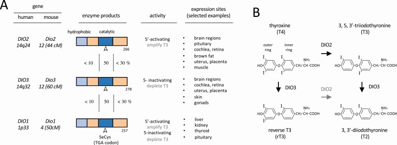
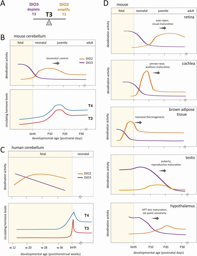
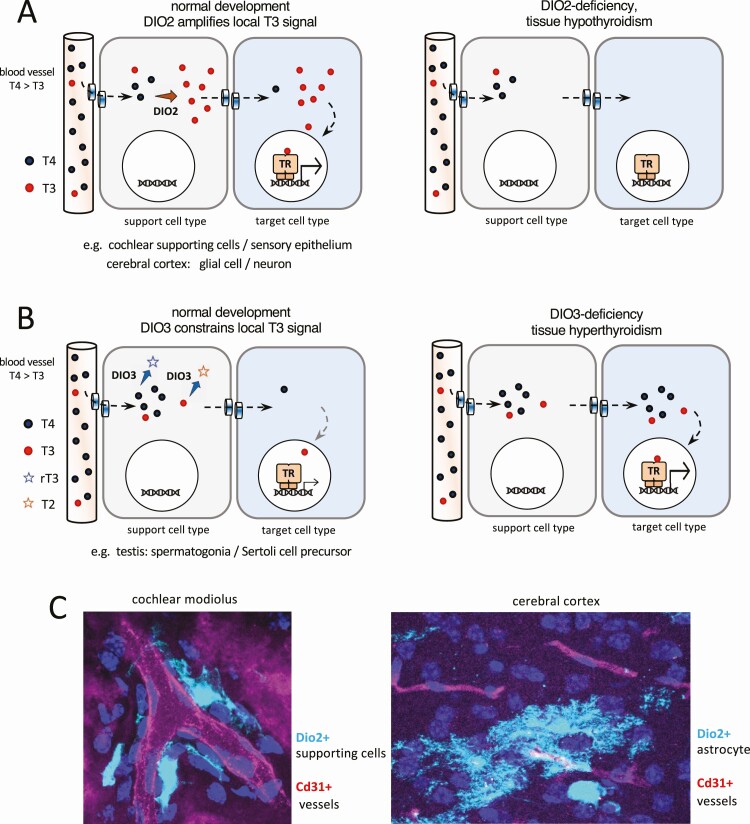
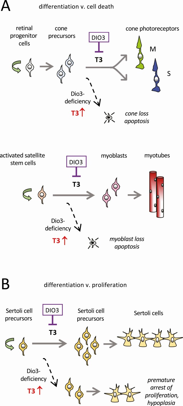
Thyroid hormones exert pleiotropic, essential actions in mammalian, including human, development. These actions depend on provision of thyroid hormones in the circulation but also to a remarkable extent on deiodinase enzymes in target tissues that amplify or deplete the local concentration of the primary active form of the hormone T3 (3,5,3'-triiodothyronine), the high affinity ligand for thyroid hormone receptors. Genetic analyses in mice have revealed key roles for activating (DIO2) and inactivating (DIO3) deiodinases in cell differentiation fates and tissue maturation, ultimately promoting neonatal viability, growth, fertility, brain development, and behavior, as well as metabolic, endocrine, and sensory functions. An emerging paradigm is how the opposing activities of DIO2 and DIO3 are coordinated, providing a dynamic switch that controls the developmental timing of a tissue response, often during neonatal and maturational transitions. A second paradigm is how cell to cell communication within a tissue determines the response to T3. Deiodinases in specific cell types, often strategically located near to blood vessels that convey thyroid hormones into the tissue, can regulate neighboring cell types, suggesting a paracrine-like layer of control of T3 action. We discuss deiodinases as switches for developmental transitions and their potential to influence tissue dysfunction in human thyroid disorders.

Keywords: central nervous system; deiodinase; development; neuroendocrine function; sensory system; thyroid hormone.


Figures

Figure 1.
Figure 1.
Main characteristics of deiodinases (DIOs). A, Protein domains of deiodinases with conservation between enzyme domains indicated, type of activity, and selected main sites of expression. B, Structure of thyroid hormones and main biochemical conversions catalyzed by the type 2 deiodinase (DIO2) and type 3 deiodinase (DIO3) enzymes (bold arrows).
Figure 2.
Figure 2.
Type 3 deiodinase (DIO3) to type 2 deiodinase (DIO2) switches in developmental transitions. A, Opposing activities of DIO3 and DIO2 provide dynamically balanced control of the 3,5,3′-triiodothyronine (T3) signal in a tissue. B and C, DIO3 to DIO2 switches in cerebellar development in mice and humans, and relationship to systemic thyroxine (T4) and T3 levels. (Cerebellar differentiation and the increase in circulating T4 and T3 occur earlier in human than mouse development.) The mouse profile parallels that originally shown for rat (6). The human profile is based on fetal data reported by Kester et al (19). Circulating hormone profiles are adapted from reported data for mice (11) and humans (124, 125). D, DIO3-DIO2 switches are customized in each tissue and often encompass terminal differentiation events and onset of organ function. All deiodinase activity profiles in panels B, C, and D are approximate and based on publications cited in the text.
Figure 3.
Figure 3.
Conceptual diagrams of paracrine-like control of 3,5,3′-triiodothyronine (T3) action by type 2 deiodinase (DIO2) and type 3 deiodinase (DIO3) within a tissue. A, DIO2+ cell types close to capillaries take up thyroxine (T4) from the circulation, convert T4 to T3, then transfer T3 to neighboring, T3-responsive cell types. Examples are found in the cochlea and cerebrum. B, DIO3+ support cell types deplete T4 and T3, thereby constraining the T3 signal available for other response cell types. An example is found in the testis. Transplasma membrane transport of T4 and T3 by specific transporter proteins is an integral part of the models shown in panels A and B. C, Images of DIO2+ support cells (pale blue) in proximity to blood vessels (red): specifically DIO2+ supporting cells in the cochlea and DIO2+ astrocytes in the cerebral cortex (images are the authors’ own work using a Dio2cre knockin; manuscript in preparation). TR, thyroid hormone receptor in the nucleus of target cells.
Figure 4.
Figure 4.
Control of cell differentiation fates by type 3 deiodinase (DIO3). A, DIO3 critically limits the 3,5,3′-triiodothyronine (T3) signal to allow normal differentiation vs T3-induced cell death. Examples show differentiation of cone photoreceptors in the retina and myoblasts during muscle regeneration. B, For Sertoli cell precursors in the testis, DIO3 limits the T3 signal to allow an appropriate period of proliferation vs premature arrest of proliferation, thereby generating a full complement of Sertoli cells.

References

    1. Osler W. Sporadic Cretinism in America. Transactions; Congress of American Physicians and Surgeons. 1897;4:169-206.
    1. Polak M, Legac I, Vuillard E, Guibourdenche J, Castanet M, Luton D. Congenital hyperthyroidism: the fetus as a patient. Horm Res. 2006;65(5):235-242. - PubMed
    1. Bianco AC, Salvatore D, Gereben B, Berry MJ, Larsen PR. Biochemistry, cellular and molecular biology, and physiological roles of the iodothyronine selenodeiodinases. Endocr Rev. 2002;23(1):38-89. - PubMed
    1. Galton VA, Larsen PR, Berry MJ. The deiodinases: their identification and cloning of their genes. Endocrinology. 2021;162(3):bqab005. - PMC - PubMed
    1. Bianco AC, Dumitrescu A, Gereben B, et al. Paradigms of dynamic control of thyroid hormone signaling. Endocr Rev. 2019;40(4):1000-1047. - PMC - PubMed

Publication types