

Postmortem pathological changes in extrapulmonary organs in SARS-CoV-2 rt-PCR-positive cases: a single-center experience
- PMID: 33963513
- PMCID: PMC8104039
- DOI: 10.1007/s11845-021-02638-8
Postmortem pathological changes in extrapulmonary organs in SARS-CoV-2 rt-PCR-positive cases: a single-center experience
Abstract
Background: Although the lung is seen as the main target organ affected by SARS-CoV-2, other organs are also damaged.
Aim: We aimed to determine the extrapulmonary findings of autopsies performed on cases with positive results with postmortem polymerase chain reaction test.
Methods: Pathological changes in extrapulmonary organs were examined with light microscopy.
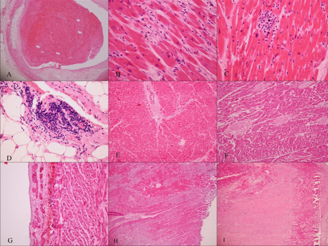
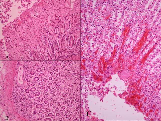
Results: Heart, liver, spleen, kidney, pancreas, and central nervous system samples of these cases were evaluated. About 80% of the cases were men, and 20% were women. In the examination of heart, 28 of the cases had scar, 14 had acute myocardial infarction, 6 had acute and previous myocardial infarction findings, 2 had myocarditis, and 4 had interstitial mononuclear inflammatory cell infiltration. In the examination of the liver, portal inflammation was observed in 84 of the cases, steatosis in 54, centrilobular necrosis in 9, and capillary endotheliitis in the portal area in 7 of them. In the evaluation of the kidney, 37 cases had chronic pyelonephritis, 36 had tubular damage, 15 had tubulointerstitial necrosis, 16 had subcapsular microhemorrhage, 10 had capillary endothelitis, and 9 had a microvascular fibrin trombosis in their glomerular capillaries. In the central nervous system, 8 cases had infarction and liquefaction, 56 had perivascular petechial hemorrhage, 54 had acute hypoxic ischemic change, 3 had parenchymal microhemorrhage, and 52 had capillary endotheliitis.
Conclusion: Autopsies play an important role in systematically examining the damage caused by the virus in all organs in order to elucidate the pathogenesis of SARS-CoV-2 infection and contribute to the clinical management of infected patients.
Keywords: Autopsy; COVID-19; Endotheliitis; Pathology; Postmortem; SARS-CoV-2; Thrombosis.
© 2021. Royal Academy of Medicine in Ireland.
Figures


References
-
- WHO Coronavirus Disease Dashboard. https://covid19.who.int/. Accessed 1 Feb 2021
-
- Republic of Turkey Ministry of Health. Covid-19 Information Page. https://covid19.saglik.gov.tr/. Accessed 1 Feb 2021
-
- de Groot RJ, Baker S, Baric R et al (2012) Family coronaviridae. In: King AMQ, Adams MJ, Cartens EB, Le owitz EJ (eds.), Virus taxonomy, the 9th report of the International Committee on Taxonomy of Viruses. Academic Press, San Diego, CA. 10.1016/B978-0-12-384684-6.00068-9 p. 806–828
MeSH terms
LinkOut - more resources
Full Text Sources
Other Literature Sources
Medical
Miscellaneous
