. 2021 Jul 2;49(W1):W359-W365.
doi: 10.1093/nar/gkab336.

LZerD webserver for pairwise and multiple protein-protein docking

Affiliations

LZerD webserver for pairwise and multiple protein-protein docking

Charles Christoffer et al. Nucleic Acids Res. .

Abstract

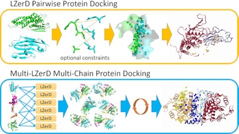
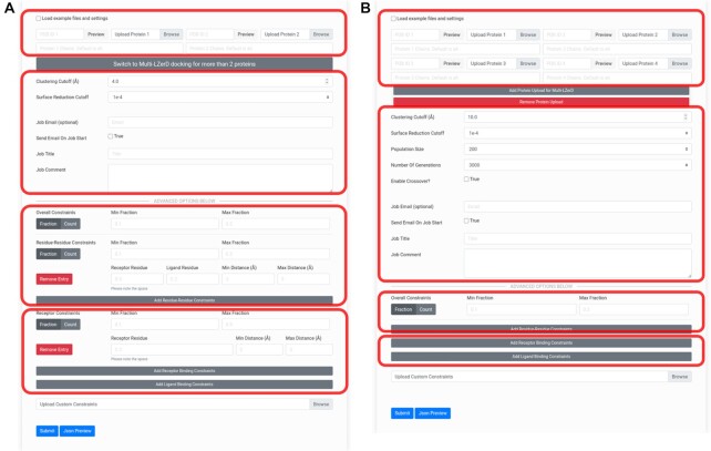
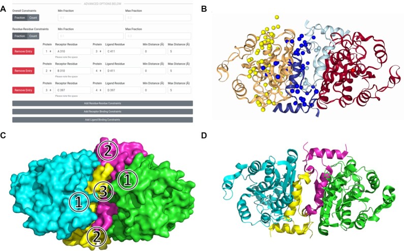
Protein complexes are involved in many important processes in living cells. To understand the mechanisms of these processes, it is necessary to solve the 3D structures of the protein complexes. When protein complex structures have not yet been determined by experiment, protein-protein docking tools can be used to computationally model the structures of these complexes. Here, we present a webserver which provides access to LZerD and Multi-LZerD protein docking tools. The protocol provided by the server have performed consistently among the top in the CAPRI blind evaluation. LZerD docks pairs of structures, while Multi-LZerD can dock three or more structures simultaneously. LZerD uses a soft protein surface representation with 3D Zernike descriptors and explores the binding pose space using geometric hashing. Multi-LZerD performs multi-chain docking by combining pairwise solutions by LZerD. Both methods output full-atom docked models of the input proteins. Users can also input distance constraints between interacting or non-interacting residues as well as residues that locate at the interface or far from the interface. The webserver is equipped with a user-friendly panel that visualizes the distribution and structures of binding poses of top scoring models. The LZerD webserver is available at https://lzerd.kiharalab.org.


Figures

Graphical Abstract
Graphical Abstract
The LZerD Protein Docking Server provides pairwise docking with LZerD and multi-chain docking with Multi-LZerD.
Figure 1.
Figure 1.
Docking job submission interfaces. (A) The submission form for pairwise LZerD protein–protein docking jobs. The first area outlined in red is for specifying input subunit structures: the toggle allows users to quickly load example subunit structures and constraints, while the file upload buttons allow users to upload their own structures. Users can alternatively specify an entry to fetch directly from the PDB. Chain IDs to extract from the individual subunits can be entered. When a structure is loaded, a 3D interactive preview is displayed. Then, there is a button to switch to the Multi-LZerD form. The second area is for specifying general job parameters: the clustering RMSD cutoff controls the structural redundancy of the output models (higher RMSD means more diverse output structures), the surface reduction cutoff controls the fineness of the conformational sampling (lower means finer sampling), and optional user email and job title and comment settings control notifications and annotations. In the third and fourth areas, in the Advanced Options section, residue-residue or binding site constraints, respectively, can be added by clicking the Add buttons and filling in the residue number(s) and distance range fields. Users can toggle between min/max fraction and min/max count of constraints that must be satisfied. For more details, check the How to Use page on the server. (B) The submission form for Multi-LZerD multiple docking jobs involving 3–6 proteins. The Multi-LZerD form has Add and Remove buttons below the file upload buttons, which can be used to add or remove subunit structures. Any number of subunit structures from 3 to 6 may be uploaded. The areas outlined in red correspond to the outlined areas of the LZerD submission form.
Figure 2.
Figure 2.
Docking input and results for HopQ-II:CEACAM1 pairwise docking. The template-based models for HopQ-II and CEACAM1 are shown in red and blue respectively in their best-scoring docked conformations. The native complex structure is shown likewise in green and cyan. (A) The constraints section of the LZerD submission form with the receptor and ligand binding sites for the constrained docking filled in. Here, receptor model chain A residue 249 is specified to be in the range of 0 to 5 Å from the nearest ligand heavy atom. Also, ligand model chain B residue 26 is specified to be in the range of 0 to 5 Å from the nearest receptor heavy atom. The min/max fraction fields are blank, leaving the default that all constraints must be satisfied simultaneously. (B) The results of docking without constraints. The centroids of the top 50 models by ranksum are shown as orange spheres. The top model shown has an I-RMSD of 3.1 Å and an formula image of 0.39, which is considered of acceptable quality under the CAPRI criteria. (C) The results of docking with the constraints shown in A. the centroids of the top 50 models by ranksum are shown as orange spheres. The top model shown has an I-RMSD of 2.6 Å and an formula image of 0.41, which is considered of medium quality under the CAPRI criteria. (D) The native structure of the complex (PDB ID: 6GBH), shown with the receptor (HopQ-II) superimposed to the same orientation as in the docked models.
Figure 3.
Figure 3.
Docking input and results for enoyl-ACP reductase multiple docking. This complex has four chains of size between 60 and 229 amino acids. (A) The constraints section of the Multi-LZerD submission form with the residue-residue interaction information to be integrated filled in. Here, distances in the range of 0 to 5 Å are specified for each of the pairs: subunit 1 chain A residue 310 and subunit 3 chain C residue 411, subunit 2 chain B residue 310 and subunit 4 chain D residue 411, and subunit 3 chain C residue 397 and subunit 4 chain D residue 397. The min/max fraction fields are blank, leaving the default that all constraints must be satisfied simultaneously. (B) The results of docking. The centroids of the top 50 models by ranksum are shown as spheres and are colored according to which subunit they represent. (C) A diagram of the protein-protein interfaces. In the top model shown in B, formula image
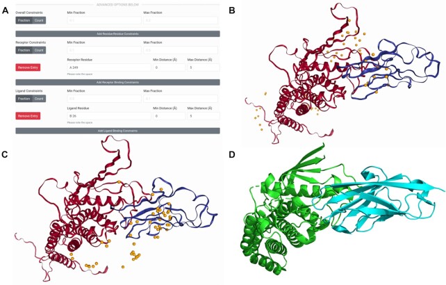
has an I-RMSD of 1.05 Å and an formula image of 0.79, formula imagehas an I-RMSD of 1.38 Å and an formula image of 0.91, and formula imagehas an I-RMSD of 1.02 and an formula image of 0.80. All three unique interfaces are modeled to medium quality under the CAPRI criteria. (D) The native structure of the complex (PDB 1NNU), shown superimposed to the same orientation as the docked models.

References

    1. Aderinwale T., Christoffer C.W., Sarkar D., Alnabati E., Kihara D.. Computational structure modeling for diverse categories of macromolecular interactions. Curr. Opin. Struct. Biol. 2020; 64:1–8. - PMC - PubMed
    1. Venkatraman V., Yang Y.D., Sael L., Kihara D.. Protein-protein docking using region-based 3D Zernike descriptors. BMC Bioinformatics. 2009; 10:407. - PMC - PubMed
    1. Esquivel-Rodriguez J., Yang Y.D., Kihara D.. Multi-LZerD: multiple protein docking for asymmetric complexes. Proteins. 2012; 80:1818–1833. - PMC - PubMed
    1. Mintseris J., Pierce B., Wiehe K., Anderson R., Chen R., Weng Z.. Integrating statistical pair potentials into protein complex prediction. Proteins. 2007; 69:511–520. - PubMed
    1. Dominguez C., Boelens R., Bonvin A.M.. HADDOCK: a protein-protein docking approach based on biochemical or biophysical information. J. Am. Chem. Soc. 2003; 125:1731–1737. - PubMed

Publication types

MeSH terms

LinkOut - more resources