. 2021 Jun 10;184(12):3163-3177.e21.
doi: 10.1016/j.cell.2021.04.016. Epub 2021 May 7.

Neutrophil elastase selectively kills cancer cells and attenuates tumorigenesis

Affiliations

Neutrophil elastase selectively kills cancer cells and attenuates tumorigenesis

Chang Cui et al. Cell. .

Abstract

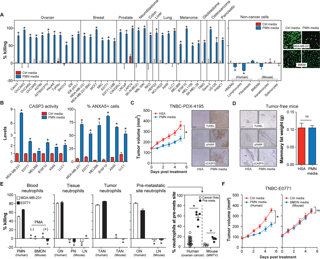
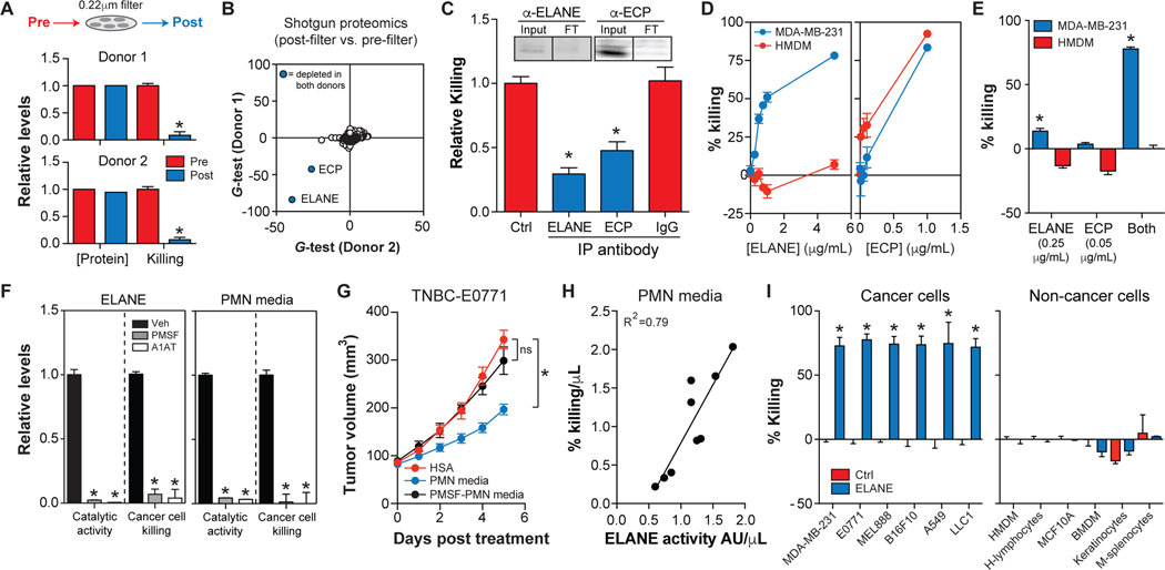
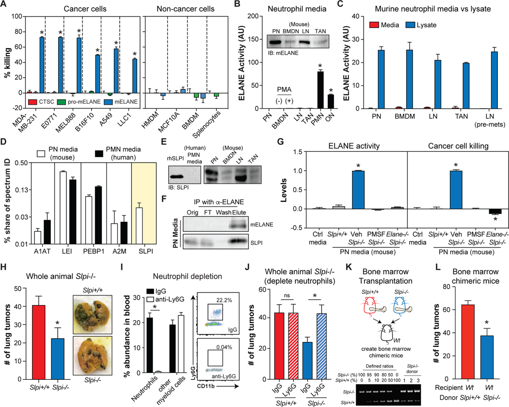
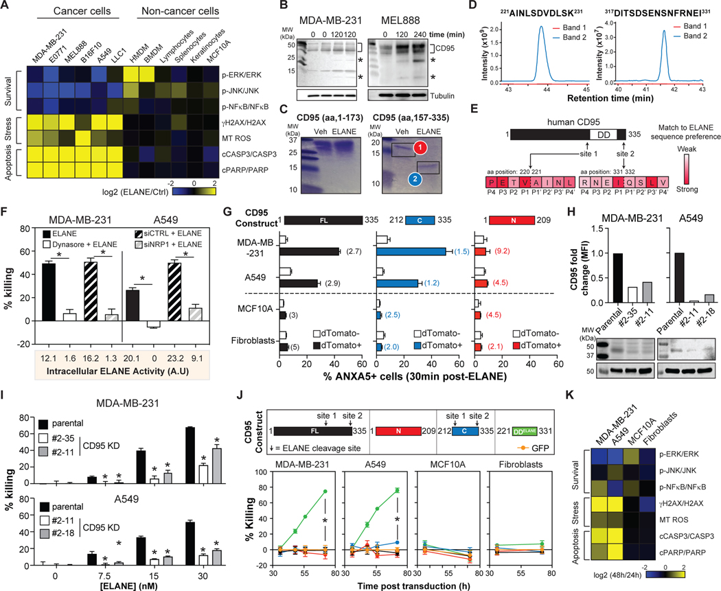
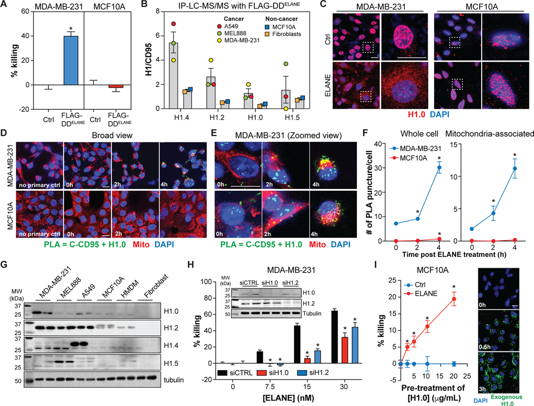
Cancer cell genetic variability and similarity to host cells have stymied development of broad anti-cancer therapeutics. Our innate immune system evolved to clear genetically diverse pathogens and limit host toxicity; however, whether/how innate immunity can produce similar effects in cancer is unknown. Here, we show that human, but not murine, neutrophils release catalytically active neutrophil elastase (ELANE) to kill many cancer cell types while sparing non-cancer cells. ELANE proteolytically liberates the CD95 death domain, which interacts with histone H1 isoforms to selectively eradicate cancer cells. ELANE attenuates primary tumor growth and produces a CD8+T cell-mediated abscopal effect to attack distant metastases. Porcine pancreatic elastase (ELANE homolog) resists tumor-derived protease inhibitors and exhibits markedly improved therapeutic efficacy. Altogether, our studies suggest that ELANE kills genetically diverse cancer cells with minimal toxicity to non-cancer cells, raising the possibility of developing it as a broad anti-cancer therapy.

Keywords: CD95; cancer; histone H1; neutrophil estate; neutrophils; therapeutics; tumor immunology.


Conflict of interest statement

Declaration of interests L.B. and C.C. are inventors on the following patent applications: UC provisional applications 62/610,711 and 62/520,325, PCT/US18/37800 and associated national filings; and US provisional application 62/782,690 and PCT/US19/67890; each filed by the University of Chicago. L.B. is an inventor on provisional patent application 63/067,059, filed by Onchilles Pharma. L.B. is a co-founder of Onchilles Pharma, a company seeking to develop ELANE/PPE-based cancer therapeutics.

Figures

Figure. 1.
Figure. 1.. Human, but not murine, neutrophils release factor(s) that selectively kill cancer cells
(A-B) Cancer or non-cancer cells were treated with PMN media (30μg/mL protein) for 24h. Cell viability was quantified by calcein-AM fluorescence and representative images are provided (A). CASP3 activity assay and ANXA5 staining (B). n=3–6/group. Scale bar = 30μm. (C) PMN media or human serum albumin (HSA) (50μg protein/day, 5 days) were injected into TNBC PDX-4195 tumors and tumor volume and apoptosis were assessed. n=14–16/group. Scale bar = 100μm. (D) PMN media or HSA (50μg protein/day, 5 days) were injected into mammary fat pads of tumor-free C57BL/6 mice. Mammary fat apoptosis and weight were monitored. n=4/group. (E) Human and murine neutrophils were isolated from various tissues. MDA-MB-231 and E0771 cell viability (calcein-AM) was assessed following treatment with neutrophil media for 24h. n=3–6/group. Neutrophil accumulation at pre-metastatic (pre-mets) sites was quantified by flow cytometry. n=5–24/group. See also Figure S1. (F) PMN media or BMDN media (50μg protein/day, 5 days) or Ctrl media were injected intratumorally into TNBC-E0771 tumors and tumor volume was monitored daily. n=7–12/group. *, p<0.05 Student’s t-test, data are mean ± SEM. Ctrl media = serum-free DMEM.
Figure. 2.
Figure. 2.. ELANE is the major anti-cancer protein in human PMN media
(A) Effect of passing PMN media through a 0.22μm filter on protein levels and MDA-MB-231 cell viability (calcein-AM). n=3/group. (B) Proteomics analysis of PMN media pre- and post-filtration. Significantly down-regulated proteins were identified by the G-test (blue circle, p<0.05 with Bonferonni correction). See also Table S2. (C) Effect of immunodepleting ECP or ELANE from PMN media on MDA-MB-231 cell viability (calcein-AM). Depletion of ECP or ELANE was confirmed by western blotting (Input = pre-depletion, FT = post-depletion). n=4/group. (D) Dose-response effect of purified human ELANE or ECP on MDA-MB-231 cell or HMDM viability (calcein-AM). n=3/group. (E) Effect of purified human ELANE or ECP on MDA-MB-231 cell or HMDM viability (calcein-AM) alone or in combination. n=3/group. (F) Effect of serine protease inhibitors (1mM PMSF or 42nM A1AT) on catalytic activity and MDA-MB-231 cancer cell viability (calcein-AM) of purified ELANE or PMN media. n=4/group. Veh = ethanol. (G) PMN media, PMSF-inactivated PMN media, or HSA were injected intratumorally into E0771 tumors and tumor volume was measured. n=8–9/group. (H) Linear regression analysis of MDA-MB-231 cell killing by PMN media vs. ELANE catalytic activity in PMN media from 9 healthy donors. n=6/donor. (I) Effect of ELANE (3μg/mL, 6h) on cancer and non-cancer cell viability (calcein-AM). n=3/group. *, p<0.05 Student’s t-test, data are mean ± SEM.
Figure. 3.
Figure. 3.. Restoring extracellular ELANE activity in murine neutrophils attenuates tumorigenesis
(A) Effect of CTSC, pro-mELANE, or mELANE (3μg/mL, 24h) on cancer and non-cancer cell viability (calcein-AM). n=6/group. (B) ELANE catalytic activity in murine and human neutrophil media. n=4/group. Immunoblotting detects mELANE in murine neutrophil media (inset). (C) Comparison of ELANE catalytic activity in murine neutrophil media (n=4/group) and lysates (n=2/group) isolated from various sources. (D) Serine protease inhibitor levels in human PMN and murine PN media quantified by shotgun proteomics, n=2/group. See also Table S3. (E) Immunoblots of SLPI in human and murine neutrophil media. Recombinant human SLPI (rhSLPI) was included as a control. (F) Co-immunoprecipitation of SLPI with mELANE in murine PN media. (G) mELANE activity in PN media from Slpi+/+, Slpi−/− mice with or without PMSF treatment, or Slpi−/− Elane−/− mice (left). Effects of those media on MDA-MB-231 cell viability (calcein-AM) (right). n=3/group. (H-J) B16F10 lung tumor colonization in whole animal Slpi−/− mice and Slpi+/+ control mice (left), along with representative images (right); n=6–10/group (H). Effect of neutrophil depletion (I) on lung tumor colonization in Slpi+/+ and Slpi−/− mice (J); n=7–11/group. (K) Schematic for generating Slpi+/+ or Slpi−/− chimeric mice (top). Engraftment efficiency was quantified using defined mixtures of Slpi+/+ and Slpi−/− bone marrow cells (bottom). (L) B16F10 lung tumor colonization in Slpi+/+ and Slpi−/− chimeric mice; n=11/group. *, p<0.05 Student’s t-test, data are mean ± SEM.
Figure. 4.
Figure. 4.. ELANE cleaves CD95 to selectively kill cancer cells
(A) Heatmap of ELANE’s (3μg/mL) effects on survival, stress, and apoptosis pathways in cancer and non-cancer cells. See also Figure S3 for raw data and quantification. (B) Effect of ELANE (3μg/mL) on CD95 cleavage in cancer cells assessed by immunoblotting using anti-C-terminal CD95 antibody. *, CD95 fragment. (C) Cleavage of the N-terminal or C-terminal domains of recombinant human CD95 by ELANE was assessed by SDS-PAGE and Coomassie blue staining. Veh = PBS. (D) Bands from (C) were analyzed by mass spectrometry to identify ELANE cleavage sites (ie. non-tryptic peptides). (E) Schematic of ELANE cleavage sites in human CD95. Heatmap shows overlap to ELANE’s sequence specificity (https://www.ebi.ac.uk/merops). (F) Effect of Dynasore (60μM, 30min), control siRNA (siCTRL) or NRP1 siRNA (siNRP1) on MDA-MB-231 and A549 cell viability (calcein) following treatment with ELANE (1.2μg/mL, 4h); n=6/group. ELANE uptake was quantified by catalytic activity in cell lysates 30min post ELANE treatment and presented below. (G) Cells transduced with various human CD95 constructs (with dTomato) were treated with ELANE (3μg/mL, 30min). Apoptosis was quantified by ANXA5 staining on dTomato+ and dTomato- populations. Flow cytometric analysis of CD95 expression levels in dTomato+ versus dTomato- cells 48h post-transduction (fold-change in parentheses). n=2/group. (H) CRISPR knockdown (KD) of CD95 in MDA-MB-231 and A549 cells was measured by flow cytometry (top) and immunoblotting (bottom). (I) Effects of ELANE (6h) on parental or CD95 KD colony viability (calcein-AM); n=6/group (J) Cells were transduced to express various human CD95 constructs or GFP (top). Cell viability was determined by calcein-AM; n=10/group (bottom). See also Figures S5A–B. (K) Heatmap of the effects DDELANE expression on survival, stress, and apoptosis pathways in cancer and non-cancer cells. See also Figures S5 for raw data and quantification. *, p<0.05 Student’s t-test, data are mean ± SEM.
Figure 5.
Figure 5.. CD95-DDELANE interacts with histone H1 to selectively kill cancer cells
(A) Effect of FLAG-DDELANE transduction on cancer and non-cancer cell viability (calcein-AM) 56h post-transduction; n=6/group (A). (B) Proteomic quantification of H1 isoform levels in anti-FLAG co-immunoprecipitations (36h post-transduction). See also Table S4. (C) Immunofluorescence of H1.0 (red) in MDA-MB-231 and MCF10A cells. Blue = DAPI. (D-E) Proximity ligation assay (PLA) of CD95 and H1.0 in MDA-MB-231 and MCF10A following treatment with ELANE (1.5μg/mL). Representative images are shown at low (D) and high (E) magnification. (F) Quantification of PLA signals in whole cells (left) and mitochondria (right). n=100 cells/group. (G) Immunoblotting of histone H1 isoforms in cancer and non-cancer cells. (H) Effects of control (siCTRL), H1.0 (siH1.0), or H1.2 (siH1.2) siRNA on MDA-MB-231 cell viability (calcein-AM) following treatment with ELANE (1.5μg/mL, 6h); n=6/group. Inset: Immunoblots confirm H1.0 and H1.2 knockdown. (I) Effects Alexa Fluor488-labeled histone H1.0 pre-treatment (3h) on MCF10A cell viability (calcein-AM) following ELANE treatment (1.5μg/mL, 18h). n=6/group. Representative images of H1.0 uptake (20μg/mL) at various time points (right). *, p<0.05 Student’s t-test, data are mean ± SEM. Scale bars = 20μm.
Figure 6.
Figure 6.. ELANE attenuates tumor growth and induces a CD8+ T cell-mediated abscopal effect
(A) E0771 tumor growth following intertumoral injection of ELANE (left, dose/day, 5 days, n=3–5/group) or HSA, PMSF-ELANE, or ELANE (middle, 12μg/day, 5 days; n=10–17/group). Inactivation of ELANE by PMSF was confirmed by activity assays (right, n=3/group). (B-C) PMSF-ELANE or ELANE (12 μg/day, 5 days) were injected into mammary fat pads of tumor-free C57BL/6 mice. Mammary fat apoptosis markers (left, B) and weight (middle, B), plasma ALT activity (right, B), and immune cell profiles (C) were measured one day after the final ELANE treatment. n=5–6/group. (D) Murine or human cancer cells were orthotopically injected into the mammary fat pad (TNBC models) or flank (melanoma and lung models). PMSF-ELANE or ELANE (12 μg/day, 5 days) were injected intratumorally and tumor volume was monitored. n=8–16/group. (E) Representative IHC images of TUNEL, cPARP, and cCASP3 staining on tumors isolated one day after the final ELANE treatment. (F-H) ELANE or PMSF-ELANE (12 μg/day, 5 days) was injected into the first palpable tumor of C3(1)-TAg mice. Tumor apoptosis (F) and immune cell composition (G) was quantified 1 day following the final ELANE treatment; n=9/group. The weight of the injected tumor (left, H), and the total weight and number of non-injected tumors (middle and right, H) were measured. n=11/group. (I) Effect of depleting CD8+ T cells on ELANE’s efficacy in injected (left) and non-injected (middle, right) tumors in the C3(1)-TAg model; n=6/group. See also Figure S6. *, p<0.05 Student’s t-test, data are mean ± SEM. Scale bars = 100μm.
Figure. 7.
Figure. 7.. PPE resists serine protease inhibition and has improved therapeutic efficacy
(A) Schematic for collecting E0771 tumor conditioned media (TCM) in serum-free DMEM for 24h. Serine protease inhibitors in the TCM were identified by immunoblotting. (B) Effects of purified A1AT (0–100nM) on ELANE and PPE (40nM) catalytic activity (n=2/group) and MDA-MB-231 cancer cell viability (calcein-AM) (n=4/group). (C) Cleavage of human CD95 N-or C-terminal recombinant proteins by ELANE or PPE was assessed by SDS-PAGE and Coomassie blue staining. (D) Effect of PPE (3μg/mL, 6h) on cancer and non-cancer cell viability (calcein-AM); n=3–6/group. (E) Effects of PPE or ELANE (0.12 units of enzyme activity) on E0771 cell viability under serum-free conditions (calcein-AM); n=3/group. (F) Effects of intratumorally injected ELANE or PPE (10 units/day for 5 and 2 days respectively) on E0771 tumor growth; n=5–6/group. (G-H) Effects of PPE (intratumoral, 10units/day, 2 days) on the injected (G) and non-injected (H) tumors in the B16F10 model with lung colonization; n=10/group. (I-J) Effects of PPE (intratumoral, 10units/day, 2 days) on the injected (I) and non-injected (J) tumors in the C3(1)-TAg model; n=10/group. (K-L) Effect of depleting CD8+ T cells on PPE’s efficacy in injected (K) and non-injected (L) tumors in the C3(1)-TAg model; n=8–9/group. See also Figure S6. *, p<0.05 Student’s t-test, data are mean ± SEM.

Comment in

Similar articles

Cited by

References

    1. Becker L, Gharib SA, Irwin AD, Wijsman E, Vaisar T, Oram JF, and Heinecke JW (2010). A macrophage sterol-responsive network linked to atherogenesis. Cell Metab. 11, 125–135. - PMC - PubMed
    1. Bode W, Meyer E, and Powers JC (1989). Human leukocyte and porcine pancreatic elastase: X-ray crystal structures, mechanism, substrate specificity, and mechanism-based inhibitors. Biochemistry 28, 1951–1963. - PubMed
    1. Catalfamo M, and Henkart PA (2003). Perforin and the granule exocytosis cytotoxicity pathway. Curr. Opin. Immunol. 15, 522–527. - PubMed
    1. Chang Y-H, Lee S-H, Liao I-C, Huang S-H, Cheng H-C, and Liao P-C (2012). Secretomic analysis identifies alpha-1 antitrypsin (A1AT) as a required protein in cancer cell migration, invasion, and pericellular fibronectin assembly for facilitating lung colonization of lung adenocarcinoma cells. Mol. Cell Proteomics 11, 1320–1339. - PMC - PubMed
    1. Chawla A, Alatrash G, Philips AV, Qiao N, Sukhumalchandra P, Kerros C, Diaconu I, Gall V, Neal S, Peters HL, et al. (2016). Neutrophil elastase enhances antigen presentation by upregulating human leukocyte antigen class I expression on tumor cells. Cancer Immunol. Immunother. 65, 741–751. - PMC - PubMed

Publication types

MeSH terms