

Social brain networks: Resting-state and task-based connectivity in youth with and without epilepsy
- PMID: 33964273
- PMCID: PMC8941486
- DOI: 10.1016/j.neuropsychologia.2021.107882
Social brain networks: Resting-state and task-based connectivity in youth with and without epilepsy
Abstract
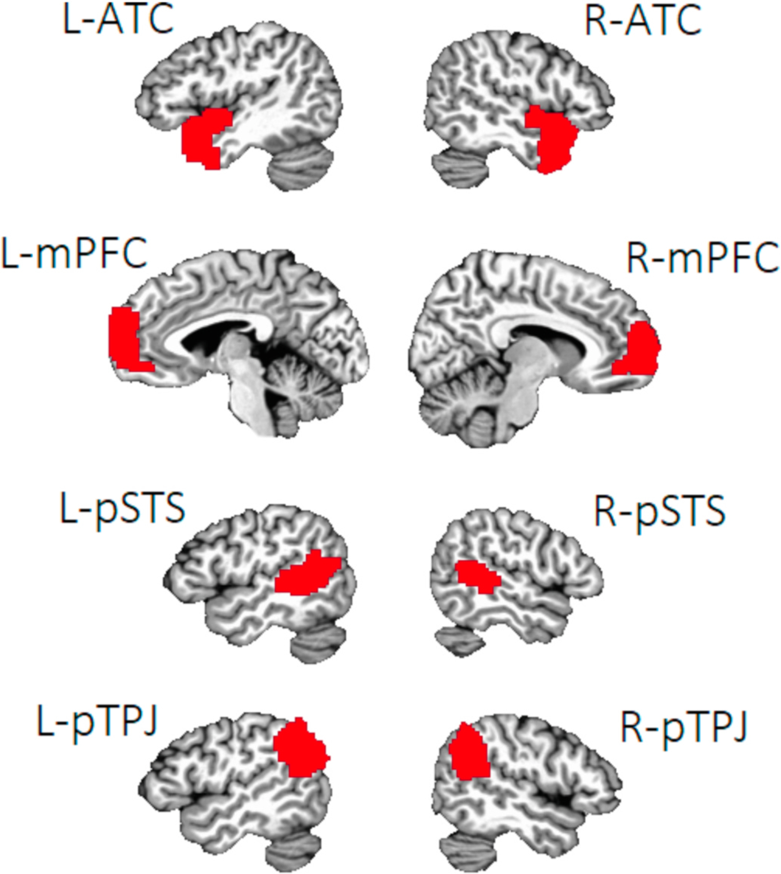
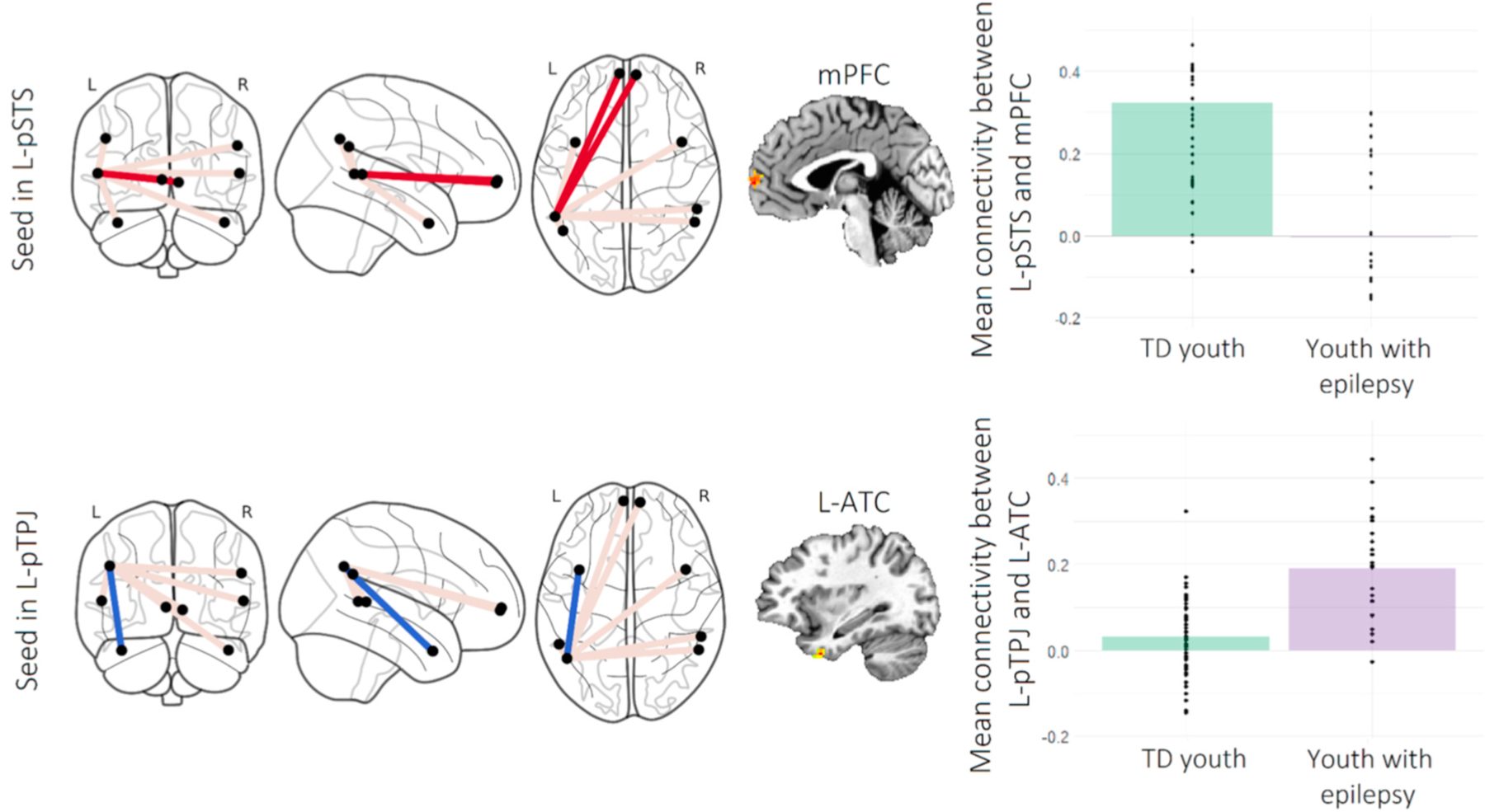
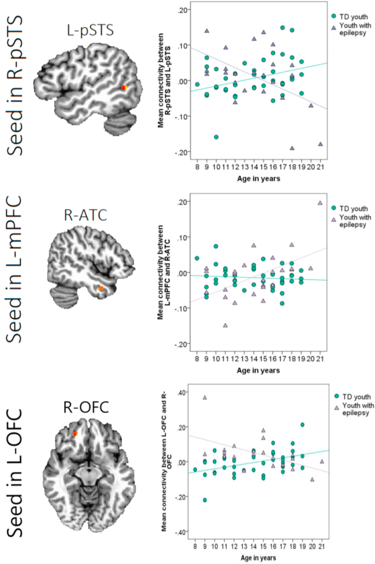
Individuals with epilepsy often experience social difficulties and deficits in social cognition. It remains unknown how disruptions to neural networks underlying such skills may contribute to this clinical phenotype. The current study compared the organization of relevant brain circuits-the "mentalizing network" and a salience-related network centered on the amygdala-in youth with and without epilepsy. Functional connectivity between the nodes of these networks was assessed, both at rest and during engagement in a social cognitive task (facial emotion recognition), using functional magnetic resonance imaging. There were no group differences in resting-state connectivity within either neural network. In contrast, youth with epilepsy showed comparatively lower connectivity between the left posterior superior temporal sulcus and the medial prefrontal cortex-but greater connectivity within the left temporal lobe-when viewing faces in the task. These findings suggest that the organization of a mentalizing network underpinning social cognition may be disrupted in youth with epilepsy, though differences in connectivity within this circuit may shift depending on task demands. Our results highlight the importance of considering functional task-based engagement of neural systems in characterizations of network dysfunction in epilepsy.
Keywords: Epilepsy; Functional connectivity; Network; Resting-state; Social brain.
Copyright © 2021 Elsevier Ltd. All rights reserved.
Figures

Similar articles
-
Linking resting-state networks and social cognition in schizophrenia and bipolar disorder.Hum Brain Mapp. 2019 Nov 1;40(16):4703-4715. doi: 10.1002/hbm.24731. Epub 2019 Jul 19. Hum Brain Mapp. 2019. PMID: 31322784 Free PMC article.
-
The neural basis of executive functioning deficits in adolescents with epilepsy: a resting-state fMRI connectivity study of working memory.Brain Imaging Behav. 2021 Feb;15(1):166-176. doi: 10.1007/s11682-019-00243-z. Brain Imaging Behav. 2021. PMID: 32043232
-
Altered functional connectivity in mesial temporal lobe epilepsy.Epilepsy Res. 2017 Nov;137:45-52. doi: 10.1016/j.eplepsyres.2017.09.001. Epub 2017 Sep 8. Epilepsy Res. 2017. PMID: 28923408
-
Applications of Resting-State Functional MR Imaging to Epilepsy.Neuroimaging Clin N Am. 2017 Nov;27(4):697-708. doi: 10.1016/j.nic.2017.06.002. Neuroimaging Clin N Am. 2017. PMID: 28985938 Review.
-
Mapping cognitive and emotional networks in neurosurgical patients using resting-state functional magnetic resonance imaging.Neurosurg Focus. 2020 Feb 1;48(2):E9. doi: 10.3171/2019.11.FOCUS19773. Neurosurg Focus. 2020. PMID: 32006946 Free PMC article. Review.
Cited by
-
Reduced eye gaze fixation during emotion recognition among patients with temporal lobe epilepsy.J Neurol. 2024 May;271(5):2560-2572. doi: 10.1007/s00415-024-12202-w. Epub 2024 Jan 30. J Neurol. 2024. PMID: 38289536
-
Artificial Intelligence in Pediatric Epilepsy Detection: Balancing Effectiveness With Ethical Considerations for Welfare.Health Sci Rep. 2025 Jan 22;8(1):e70372. doi: 10.1002/hsr2.70372. eCollection 2025 Jan. Health Sci Rep. 2025. PMID: 39846037 Free PMC article.
-
Social cognition in children and adolescents with epilepsy: A meta-analysis.Front Psychiatry. 2022 Sep 15;13:983565. doi: 10.3389/fpsyt.2022.983565. eCollection 2022. Front Psychiatry. 2022. PMID: 36186867 Free PMC article.
-
Alternative Brain Connectivity Underscores Age-Related Differences in the Processing of Interactive Biological Motion.J Neurosci. 2023 May 17;43(20):3666-3674. doi: 10.1523/JNEUROSCI.2109-22.2023. Epub 2023 Mar 24. J Neurosci. 2023. PMID: 36963845 Free PMC article.
-
Sevoflurane-induced high-frequency oscillations, effective connectivity and intraoperative classification of epileptic brain areas.Clin Neurophysiol. 2023 Jun;150:17-30. doi: 10.1016/j.clinph.2023.03.004. Epub 2023 Mar 17. Clin Neurophysiol. 2023. PMID: 36989866 Free PMC article.
References
-
- Allison T, Puce A, McCarthy G, 2000. Social perception from visual cues: role of the STS region. Trends Cognit. Sci 4 (7), 267–278. - PubMed
Publication types
MeSH terms
Grants and funding
LinkOut - more resources
Full Text Sources
Other Literature Sources
Medical
