

Primary squamous cell carcinoma of the pancreas with a large pseudocyst of the pancreas as the first manifestation: a rare case report and literature review
- PMID: 33964875
- PMCID: PMC8105924
- DOI: 10.1186/s12876-021-01804-7
Primary squamous cell carcinoma of the pancreas with a large pseudocyst of the pancreas as the first manifestation: a rare case report and literature review
Abstract
Background: Primary squamous cell carcinoma (SCC) of the pancreas with pseudocysts, especially diagnosed by endoscopic ultrasound-guided fine-needle aspiration (EUS-FNA), is extremely rare.
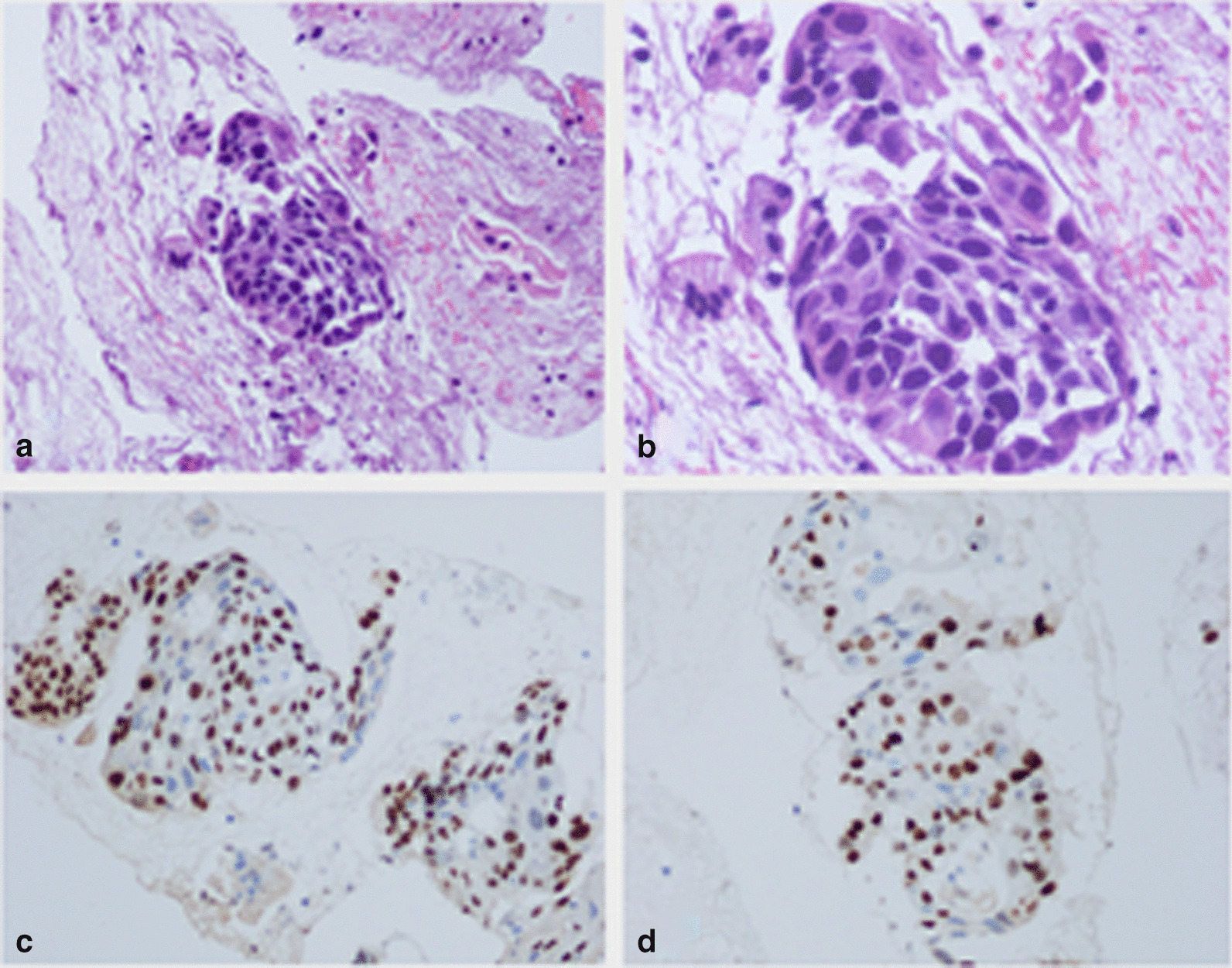
Case presentation: A 64-year-old man was admitted to our department for abdominal distension. Two months ago, he experienced abdominal pain for 1 day and was diagnosed with acute pancreatitis in another hospital. After admission, laboratory tests showed the following: amylase 400 U/L, lipase 403 U/L, and carbohydrate antigen 19-9 (CA19-9) 347 U/mL. Abdominal computed tomography (CT) revealed pancreatitis with a pseudocyst with a diameter measuring 7 cm. During linear EUS, a large pseudocyst (5.4 × 5.2 cm) was observed in the pancreatic body. EUS-FNA was performed. We obtained specimens for histopathology and placed a plastic stent through the pancreas and stomach to drain the pseudocyst. Puncture fluid examination revealed the following: CA19-9 > 12,000 U/mL carcinoembryonic antigen (CEA) 7097.42 ng/ml, amylase 27,145.3 U/L, and lipase > 6000 U/L. Cytopathology revealed an abnormal cell mass, and cancer was suspected. Furthermore, with the result of immunohistochemistry on cell mass (CK ( +), P40 ( +), p63 ( +), CK7 (-) and Ki-67 (30%)), the patient was examined as squamous cell carcinoma (SCC). However, the patient refused surgery, radiotherapy and chemotherapy. After drainage, the cyst shrank, but the patient died 3 months after diagnosis due to liver metastasis and multiple organ failure.
Conclusion: For patients with primary pancreatic pseudocysts with elevated serum CEA and CA19-9 levels, we should not rule out pancreatic cancer, which may also be a manifestation of primary pancreatic SCC. EUS-FNA is helpful for obtaining histopathology and cytology and thus improving diagnostic accuracy.
Keywords: Endoscopic ultrasound-guided fine-needle aspiration; Pancreas; Pseudocyst; Squamous cell carcinoma.
Conflict of interest statement
The authors declare that they have no competing interests.
Figures


References
-
- Attiyeh MA, Chakraborty J, Doussot A, Langdon-Embry L, Mainarich S, Gonen M, Balachandran VP, D'Angelica MI, DeMatteo RP, Jarnagin WR, et al. Survival prediction in pancreatic ductal adenocarcinoma by quantitative computed tomography image analysis. Ann Surg Oncol. 2018;25(4):1034–1042. doi: 10.1245/s10434-017-6323-3. - DOI - PMC - PubMed
-
- Tadic M, Stoos-Veic T, Kujundzic M, Turcic P, Aralica G, Boskoski I. Insulin-like growth factor 2 binding protein 3 expression on endoscopic ultrasound guided fine needle aspiration specimens in pancreatic ductal adenocarcinoma. Eur J Gastroenterol Hepatol. 2020;32(4):496–500. doi: 10.1097/MEG.0000000000001696. - DOI - PubMed
Publication types
MeSH terms
LinkOut - more resources
Full Text Sources
Other Literature Sources
Medical
Research Materials
