

A plant-based medicinal food inhibits the growth of human gastric carcinoma by reversing epithelial-mesenchymal transition via the canonical Wnt/β-catenin signaling pathway
- PMID: 33964908
- PMCID: PMC8106854
- DOI: 10.1186/s12906-021-03301-6
A plant-based medicinal food inhibits the growth of human gastric carcinoma by reversing epithelial-mesenchymal transition via the canonical Wnt/β-catenin signaling pathway
Abstract
Background: Natural products, especially those with high contents of phytochemicals, are promising alternative medicines owing to their antitumor properties and few side effects. In this study, the effects of a plant-based medicinal food (PBMF) composed of six medicinal and edible plants, namely, Coix seed, Lentinula edodes, Asparagus officinalis L., Houttuynia cordata, Dandelion, and Grifola frondosa, on gastric cancer and the underlying molecular mechanisms were investigated in vivo.
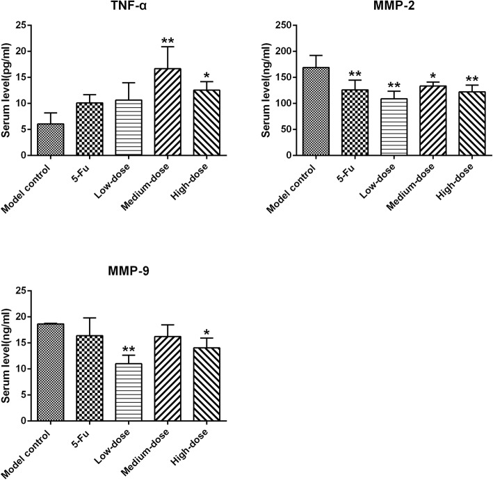
Methods: A subcutaneous xenograft model of gastric cancer was successfully established in nude mice inoculated with SGC-7901 cells. The tumor-bearing mice were separately underwent with particular diets supplemented with three doses of PBMF (43.22, 86.44, and 172.88 g/kg diet) for 30 days. Tumor volumes were recorded. Histopathological changes in and apoptosis of the xenografts were evaluated by hematoxylin and eosin staining and terminal deoxynucleotidyl transferase-mediated dUTP nick end labeling staining, respectively. Serum levels of TNF-α, MMP-2, and MMP-9 were detected by enzyme-linked immunosorbent assay. The mRNA expression levels of β-catenin, GSK-3β, E-cadherin, N-cadherin, MMP-2/9, Snail, Bax, Bcl-2, Caspase-3/9, and Cyclin D1 were evaluated via real-time quantitative polymerase chain reaction. The protein expression levels of GSK-3β, E-cadherin, N-cadherin, and Ki-67 were determined by immunohistochemistry staining.
Results: PBMF treatment efficiently suppressed neoplastic growth, induced apoptosis, and aggravated necrosis in the xenografts of SGC-7901 cells. PBMF treatment significantly decreased the serum levels of MMP-2 and MMP-9 and significantly increased that of TNF-α. Furthermore, PBMF treatment notably upregulated the mRNA expression levels of GSK-3β, E-cadherin, Bax, Caspase-3, and Caspase-9 but substantially downregulated those of β-catenin, N-cadherin, MMP-2, MMP-9, Snail, and Cyclin D1 in tumor tissues. The Bax/Bcl-2 ratio was upregulated at the mRNA level. Moreover, PBMF treatment remarkably increased the protein expression levels of GSK-3β and E-cadherin but notably reduced those of Ki-67 and N-cadherin in tumor tissues.
Conclusions: The PBMF concocted herein exerts anti-gastric cancer activities via epithelial-mesenchymal transition reversal, apoptosis induction, and proliferation inhibition. The underlying molecular mechanisms likely rely on suppressing the Wnt/β-catenin signaling pathway.
Keywords: Epithelial–mesenchymal transition; Gastric cancer; Medicinal food; Wnt/β–catenin signaling pathway.
Conflict of interest statement
The authors declare that they have no competing interests.
Figures


Similar articles
-
Effects of plant-based medicinal food on postoperative recurrence and lung metastasis of gastric cancer regulated by Wnt/β-catenin-EMT signaling pathway and VEGF-C/D-VEGFR-3 cascade in a mouse model.BMC Complement Med Ther. 2022 Sep 2;22(1):233. doi: 10.1186/s12906-022-03703-0. BMC Complement Med Ther. 2022. PMID: 36056333 Free PMC article.
-
Acetyl-11-keto-β-boswellic acid (AKBA) inhibits human gastric carcinoma growth through modulation of the Wnt/β-catenin signaling pathway.Biochim Biophys Acta. 2013 Jun;1830(6):3604-15. doi: 10.1016/j.bbagen.2013.03.003. Epub 2013 Mar 14. Biochim Biophys Acta. 2013. Retraction in: Biochim Biophys Acta. 2015 Jan;1850(1):254. doi: 10.1016/j.bbagen.2014.11.003. PMID: 23500016 Retracted.
-
Hydroalcoholic root extracts of Houttuynia cordata (Thunb.) standardized by UPLC-Q-TOF-MS/MS promotes apoptosis in human hepatocarcinoma cell HepG2 via GSK-3β/β-catenin/PDL-1 axis.Fitoterapia. 2023 Dec;171:105684. doi: 10.1016/j.fitote.2023.105684. Epub 2023 Sep 24. Fitoterapia. 2023. PMID: 37751799
-
Natural Products and Gastric Cancer: Cellular Mechanisms and Effects to Change Cancer Progression.Anticancer Agents Med Chem. 2023;23(13):1506-1518. doi: 10.2174/1871520623666230407082955. Anticancer Agents Med Chem. 2023. PMID: 37026490 Review.
-
Role of T-box transcription factor 3 in gastric cancers.World J Gastrointest Pathophysiol. 2023 Mar 22;14(2):12-20. doi: 10.4291/wjgp.v14.i2.12. World J Gastrointest Pathophysiol. 2023. PMID: 37035275 Free PMC article. Review.
Cited by
-
Houttuynia cordata Thunb: An Ethnopharmacological Review.Front Pharmacol. 2021 Sep 1;12:714694. doi: 10.3389/fphar.2021.714694. eCollection 2021. Front Pharmacol. 2021. PMID: 34539401 Free PMC article. Review.
-
Enhancement of apoptosis in HCT116 and HepG2 cells by Coix lacryma-jobi var. lacryma-jobi seed extract in combination with sorafenib.Chin Herb Med. 2025 Feb 21;17(2):322-339. doi: 10.1016/j.chmed.2025.02.005. eCollection 2025 Apr. Chin Herb Med. 2025. PMID: 40256710 Free PMC article.
-
Pharmacological role of Herba Patriniae and Coix seed in colorectal cancer.World J Gastrointest Oncol. 2025 Mar 15;17(3):99673. doi: 10.4251/wjgo.v17.i3.99673. World J Gastrointest Oncol. 2025. PMID: 40092956 Free PMC article.
-
Effects of plant-based medicinal food on postoperative recurrence and lung metastasis of gastric cancer regulated by Wnt/β-catenin-EMT signaling pathway and VEGF-C/D-VEGFR-3 cascade in a mouse model.BMC Complement Med Ther. 2022 Sep 2;22(1):233. doi: 10.1186/s12906-022-03703-0. BMC Complement Med Ther. 2022. PMID: 36056333 Free PMC article.
-
How Should the Worldwide Knowledge of Traditional Cancer Healing Be Integrated with Herbs and Mushrooms into Modern Molecular Pharmacology?Pharmaceuticals (Basel). 2022 Jul 14;15(7):868. doi: 10.3390/ph15070868. Pharmaceuticals (Basel). 2022. PMID: 35890166 Free PMC article. Review.
References
MeSH terms
Substances
LinkOut - more resources
Full Text Sources
Other Literature Sources
Medical
Research Materials
Miscellaneous
