. 2021 May 8;40(1):161.
doi: 10.1186/s13046-021-01962-2.

Interferon- and STING-independent induction of type I interferon stimulated genes during fractionated irradiation

Affiliations

Interferon- and STING-independent induction of type I interferon stimulated genes during fractionated irradiation

Ruben S A Goedegebuure et al. J Exp Clin Cancer Res. .

Abstract

Background: Improvement of radiotherapy efficacy requires better insight in the dynamic responses that occur during irradiation. Here, we aimed to identify the molecular responses that are triggered during clinically applied fractionated irradiation.

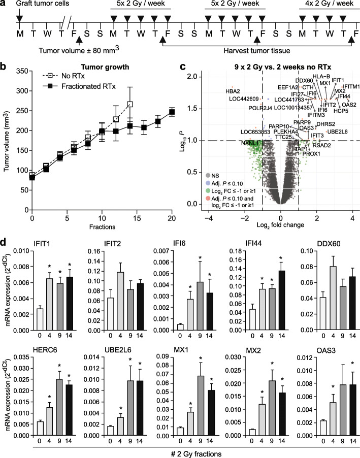
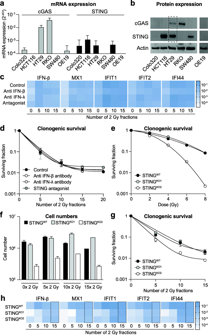
Methods: Gene expression analysis was performed by RNAseq or microarray analysis of cancer cells or xenograft tumors, respectively, subjected to 3-5 weeks of 5 × 2 Gy/week. Validation of altered gene expression was performed by qPCR and/or ELISA in multiple cancer cell lines as well as in pre- and on-treatment biopsies from esophageal cancer patients ( NCT02072720 ). Targeted protein inhibition and CRISPR/Cas-induced gene knockout was used to analyze the role of type I interferons and cGAS/STING signaling pathway in the molecular and cellular response to fractionated irradiation.

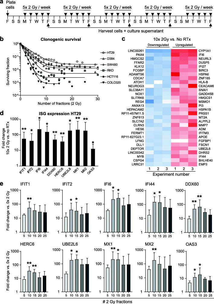
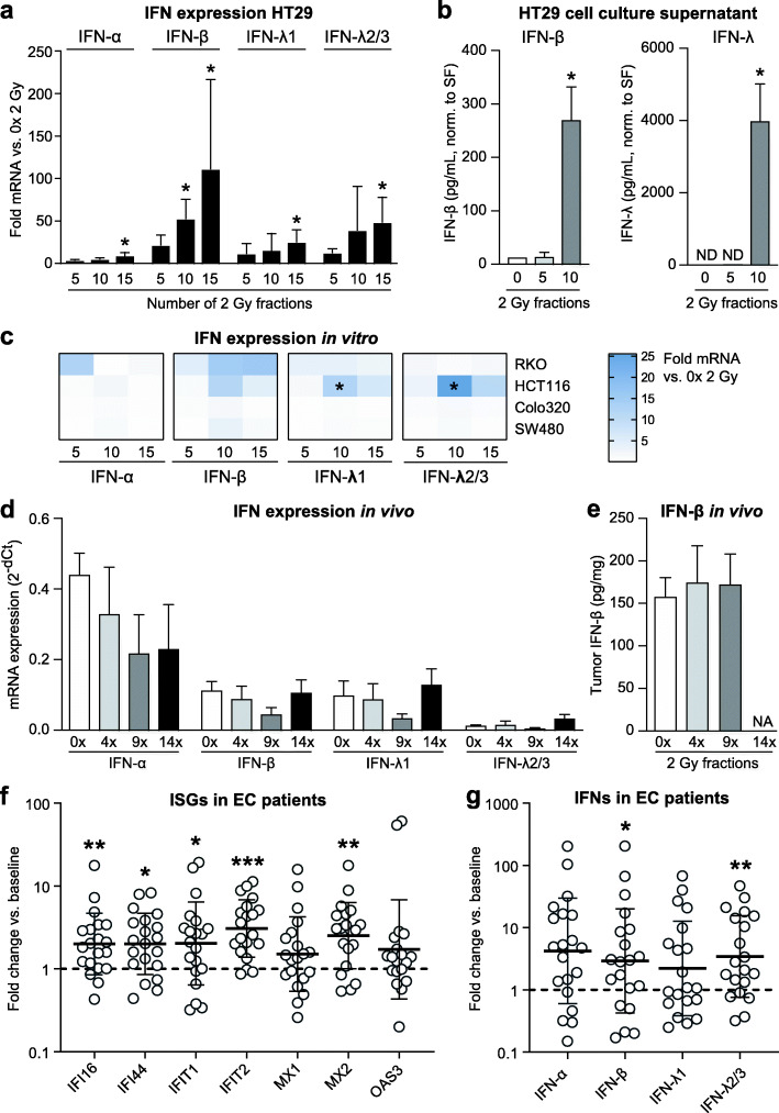
Results: Gene expression analysis identified type I interferon signaling as the most significantly enriched biological process induced during fractionated irradiation. The commonality of this response was confirmed in all irradiated cell lines, the xenograft tumors and in biopsies from esophageal cancer patients. Time-course analyses demonstrated a peak in interferon-stimulated gene (ISG) expression within 2-3 weeks of treatment. The response was accompanied by a variable induction of predominantly interferon-beta and/or -lambda, but blocking these interferons did not affect ISG expression induction. The same was true for targeted inhibition of the upstream regulatory STING protein while knockout of STING expression only delayed the ISG expression induction.

Conclusions: Collectively, the presented data show that clinically applied fractionated low-dose irradiation can induce a delayed type I interferon response that occurs independently of interferon expression or STING signaling. These findings have implications for current efforts that aim to target the type I interferon response for cancer treatment.

Keywords: Immune response; Radiotherapy; Type I interferons.


Conflict of interest statement

The authors declare that the research was conducted in the absence of any commercial or financial relationships that could be construed as a potential conflict of interest.

Figures

Fig. 1
Fig. 1
Induction of a type I IFN response in cancer cells peaks after 2 weeks of fractionated irradiation in vitro. Fractionated irradiation induces a type I IFN response in cancer cells, which peaks after 2 weeks and coincides with a convergence in clonogenic survival to a steady state. a Scheme of fractionated irradiation applied to human cancer cells in vitro. b Clonogenic survival analyses show a log-linear decline in survival during the first 2 weeks of treatment after which a steady-state survival is reached up to 6 weeks of treatment. Adapted from Van den Berg et al. [11]. c Heat map showing the 30 most downregulated and upregulated genes after 2 weeks of treatment vs. untreated as determined by RNA deep-sequencing of HT29 cells (n = 3). d The mRNA expression induction of a panel of 10 IFN-stimulated genes (ISGs) after 2 weeks of treatment was confirmed by qPCR (n = 3). Geometric mean + SD is shown. * p-value ≤0.05 vs. no radiotherapy (RTx). e Time course analysis of ISG mRNA expression induction shows a peak starting around 2 weeks of treatment (n = 3). Geometric mean + SD is shown. * p-value ≤0.05 vs 0 × 2 Gy
Fig. 2
Fig. 2
Induction of a type I IFN response in tumor tissue peaks after 2 weeks of fractionated irradiation in vivo. The induction of a type I IFN response upon fractionated radiotherapy is confirmed in a HT29 xenograft model. a Scheme of fractionated irradiation applied to HT29 xenograft tumor in mice. b Tumor growth curves of HT29 xenograft tumors with (black squares) or without (white squares) irradiation. Note the growth delay starts around day 10 and recovers around day 17 (n = 5 mice/group). c Volcano plot of microarray data comparing gene expression in HT29 xenograft tumors after 2 weeks of RTx vs. no radiotherapy (RTx). NS = not significant. FC = fold change. d Time course analysis of ISG mRNA expression induction shows a gradual increase that peaks around 2 weeks of treatment. * p-value ≤0.05 vs. 0 × 2 Gy
Fig. 3
Fig. 3
Patterns of type I and III interferon induction upon fractionated irradiation. Different patterns of either type I and/or type III interferon induction occur in vitro, in vivo and patients with esophageal cancer during the course of fractionated radiotherapy, independent of ISG induction. a mRNA expression analyses of interferon expression in HT29 cells during fractionated irradiation (n = 3). * p-value ≤0.05 vs. 0 × 2 Gy. b Levels of IFN-β and IFN-λ protein in cell culture supernatants of HT29 cells during fractionated irradiation (n = 3). * p-value ≤0.05 vs. 0 × 2 Gy. CM = culture medium. SF = surviving fraction. c mRNA expression analyses of interferon expression in RKO, HCT116, COLO320 and SW480 cells during fractionated irradiation vs. 0 × 2 Gy. * p-value ≤0.05 vs. 0 × 2 Gy. d mRNA expression analyses of interferon expression in HT29 xenograft tumors during fractionated irradiation (n = 5 mice/group). e Levels of IFN-β and IFN-λ protein in mouse serum during fractionated irradiation (n = 5 mice/group). f mRNA expression levels of ISG expression in patient-matched tumor samples from esophageal cancer patients (n = 20) prior to or during chemoradiotherapy. Fold expression in on-treatment samples vs. pre-treatment is shown. * p-value ≤0.05 vs. matched pre-treatment samples. g Similar as in (f) for fold change in mRNA expression levels of different IFNs. * p-value ≤0.05 vs. matched pre-treatment samples
Fig. 4
Fig. 4
ISG induction upon fractionated irradiation occurs independent of STING, IFN-β or IFN-λ. Interferon stimulated genes (ISGs) can be induced independent of the interferons known to mediate this response, or the upstream regulator protein STING. a mRNA expression levels of cGAS (grey bars) and STING (black bars) in different cancer cell lines. b Western blots showing protein expression of cGAS and STING in different cancer cell lines. Actin staining was used as loading control. The dotted box shows the only cell line, i.e. HT29, in which both cGAS and STING protein expression could be detected. c Heat map of mRNA expression of different ISGs and IFN-β in HT29 cells treated with fractionated irradiation in the presence or absence of either anti IFN-β antibody, anti IFN-λ antibody or a STING antagonist. No significant changes were observed in the presence of any of the treatments as compared to irradiation alone (n = 3). d Clonogenic survival of HT29 during fractionated irradiation in the presence or absence of either anti IFN-β antibody, anti IFN-λ antibody or a STING antagonist. No significant changes in surviving fractions were observed in the presence of any of the latter treatments as compared to irradiation alone (n = 3). e Clonogenic survival of HT29 wild-type cells and two HT29 STING knockout cells in response to single dose irradiation. STINGKO2 shows higher radiosensitivity as compared to wild type cells. f Cell numbers of HT29 wild-type cells and two HT29 STING knockout cells during fractionated irradiation. While STINGKO2 displayed slower growth already at base-line, fractionated irradiation did not affect growth of knockout cells compared to wild type cells. g Clonogenic survival of HT29 wild-type cells and two HT29 STING knockout cells during fractionated irradiation. STINGKO2 shows higher radiosensitivity as compared to wild type cells. h) Heat map of mRNA expression of different ISGs and IFN-β in HT29 wild-type cells and two HT29 STING knockout cells during fractionated irradiation (n = 2). At baseline (0 × 2 Gy) both knockout cell lines show lower expression of all genes analyzed as compared to wild-type cells. At the end of the treatment period (15 × 2 Gy, dotted box) no more difference in expression levels is observed in wild-type vs. knockout cells for any of the genes analyzed

References

    1. Bernier J, Hall EJ, Giaccia A. Radiation oncology: a century of achievements. Nat Rev Cancer. 2004;4(9):737–747. doi: 10.1038/nrc1451. - DOI - PubMed
    1. Baumann M, Krause M, Hill R. Exploring the role of cancer stem cells in radioresistance. Nat Rev Cancer. 2008;8(7):545–554. doi: 10.1038/nrc2419. - DOI - PubMed
    1. Begg AC, Stewart FA, Vens C. Strategies to improve radiotherapy with targeted drugs. Nat Rev Cancer. 2011;11(4):239–253. doi: 10.1038/nrc3007. - DOI - PubMed
    1. Goedegebuure RSA, de Klerk LK, Bass AJ, Derks S, Thijssen VLJL. Combining radiotherapy with anti-angiogenic therapy and immunotherapy; a therapeutic triad for cancer. Front Immunol. 2019;9:3107. doi: 10.3389/fimmu.2018.03107. - DOI - PMC - PubMed
    1. Morgan MA, Lawrence TS. Molecular pathways: overcoming radiation resistance by targeting DNA damage response pathways. Clin Cancer Res. 2015;21(13):2898–2904. doi: 10.1158/1078-0432.CCR-13-3229. - DOI - PMC - PubMed

MeSH terms