. 2021 May 8;9(1):82.
doi: 10.1186/s40478-021-01190-x.

Early onset senescence and cognitive impairment in a murine model of repeated mTBI

Affiliations

Early onset senescence and cognitive impairment in a murine model of repeated mTBI

Nicole Schwab et al. Acta Neuropathol Commun. .

Abstract

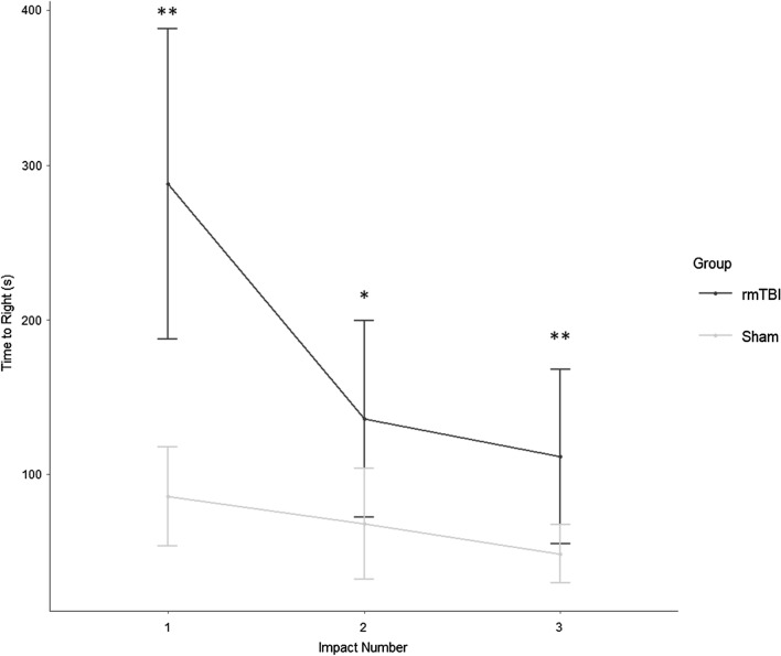
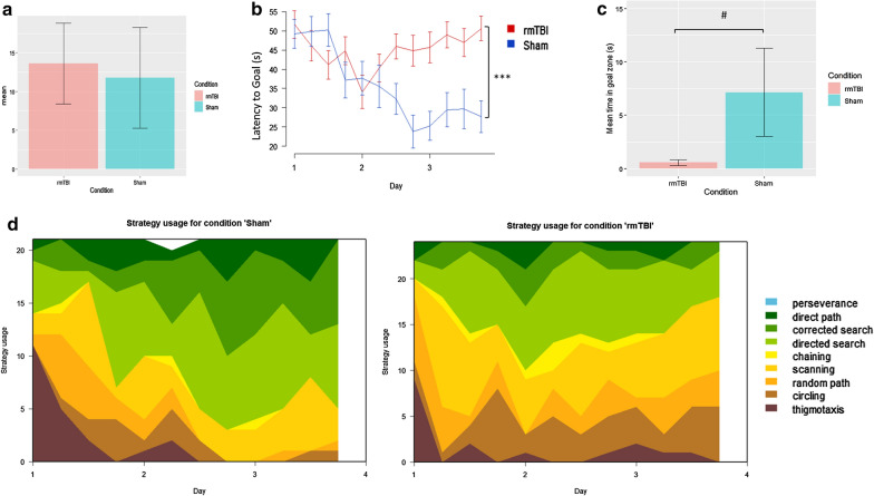
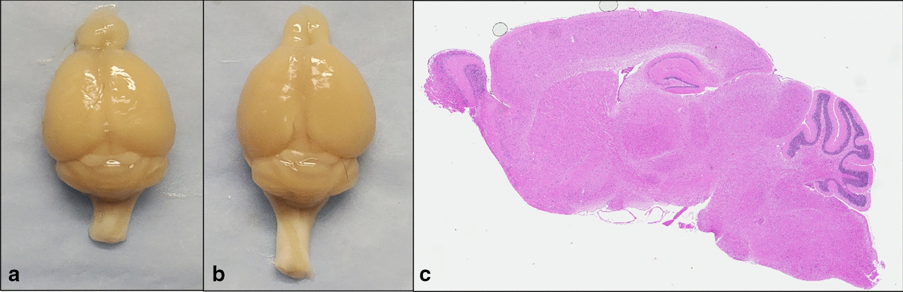
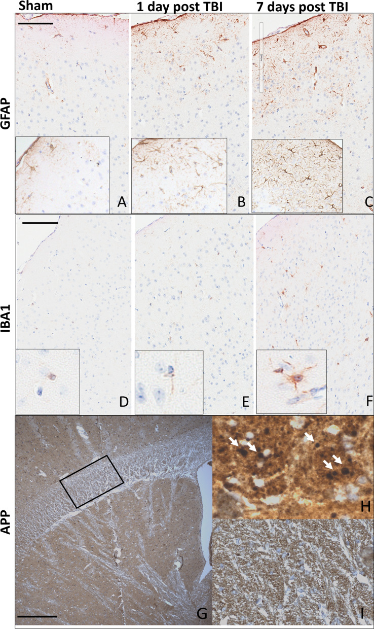
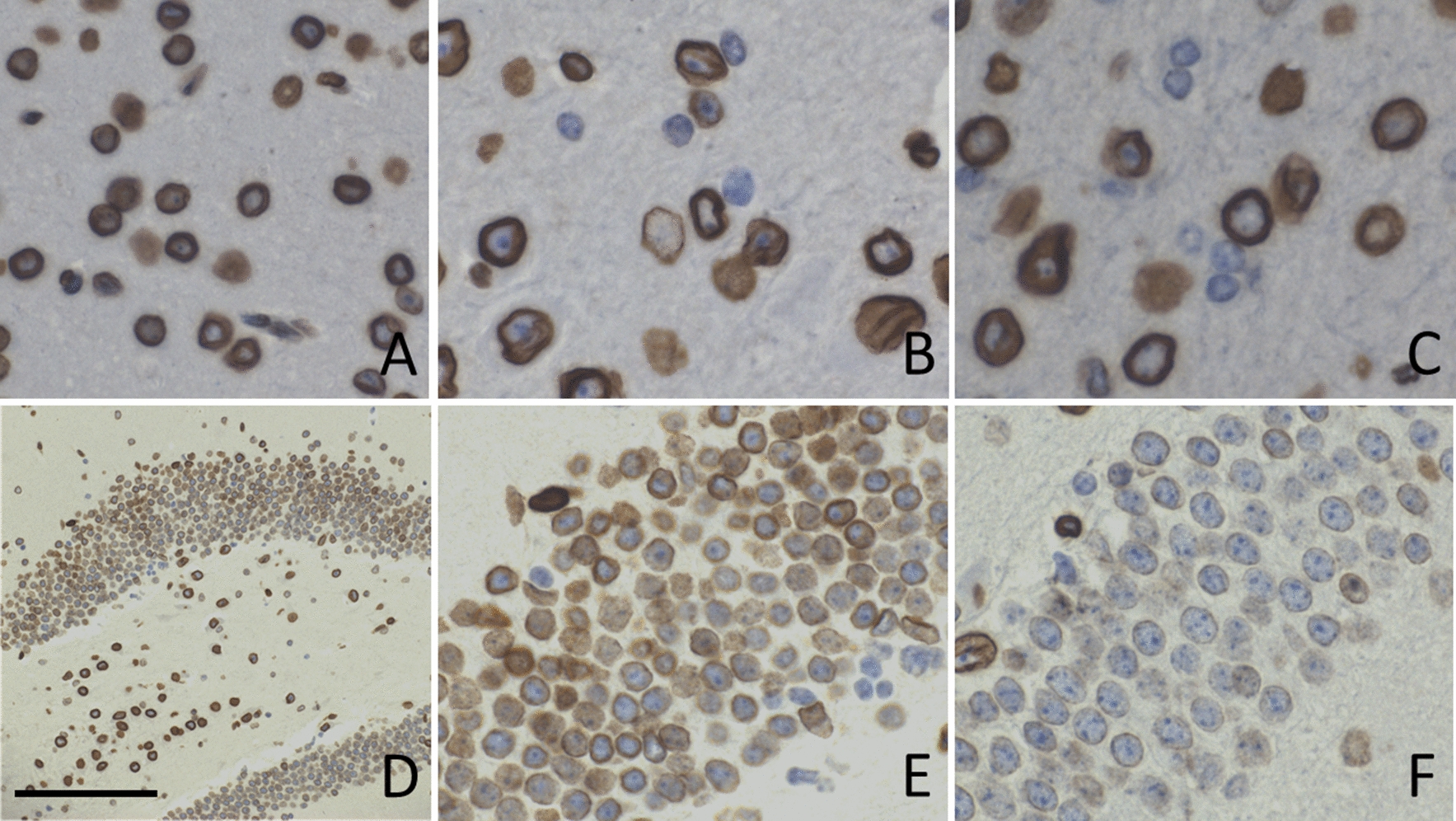
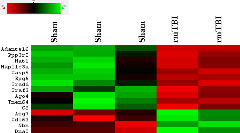
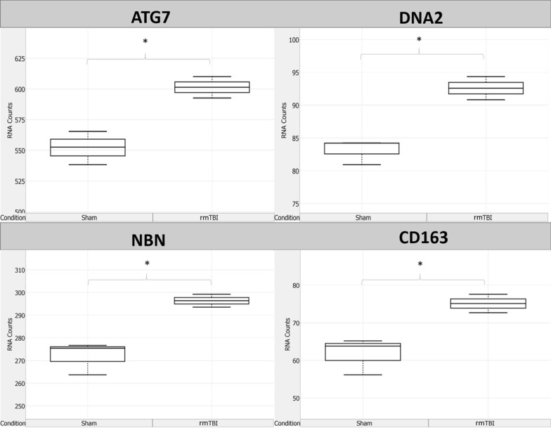
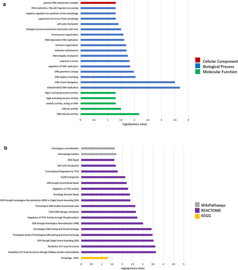
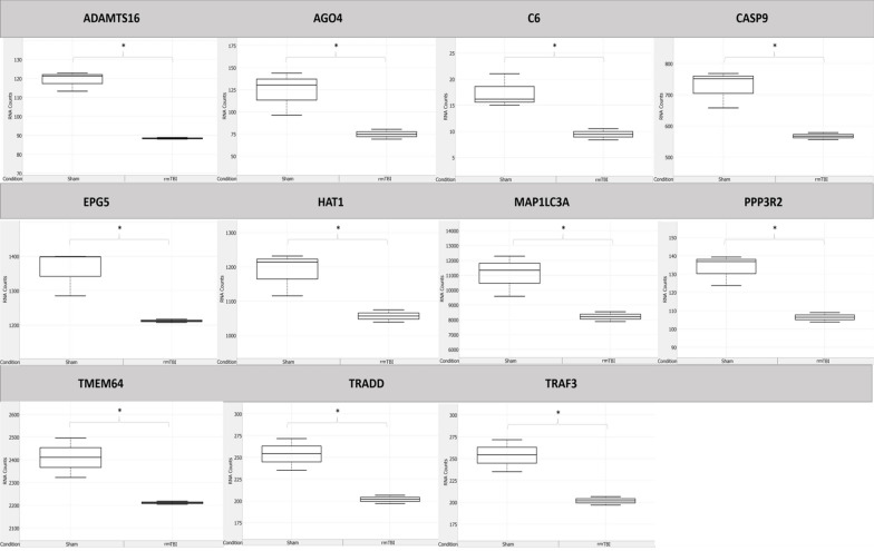
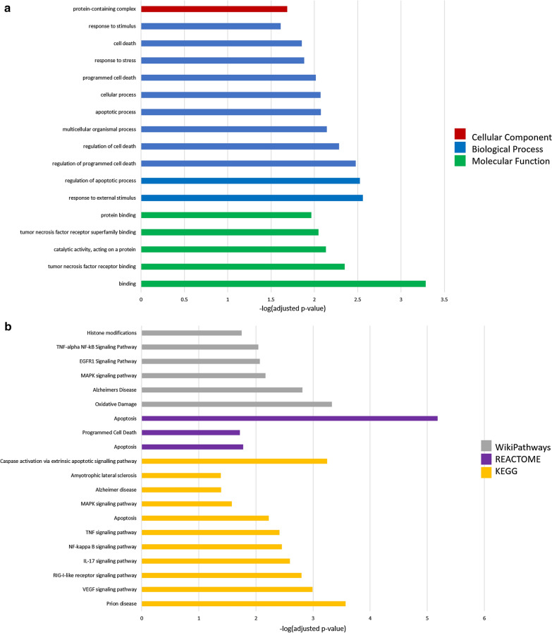
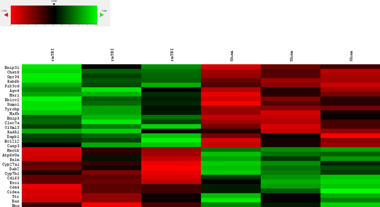
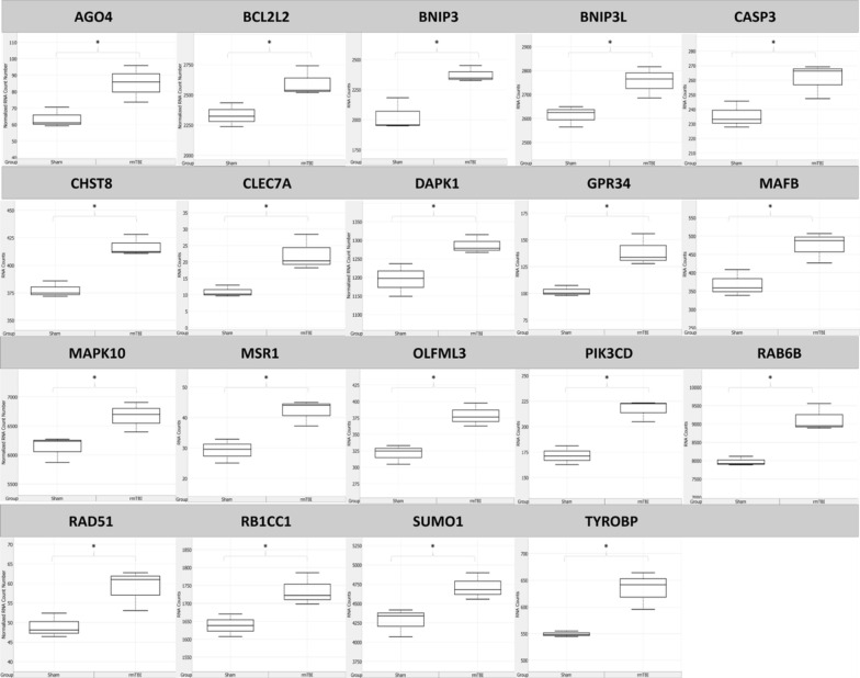
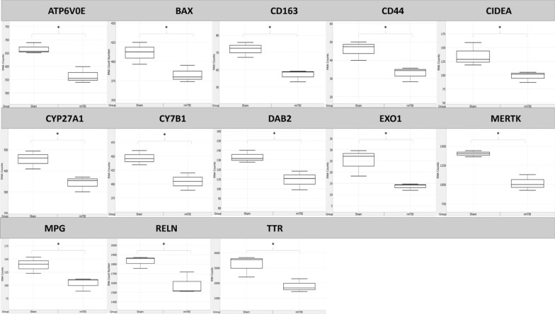
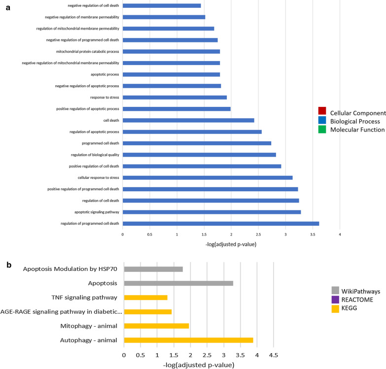
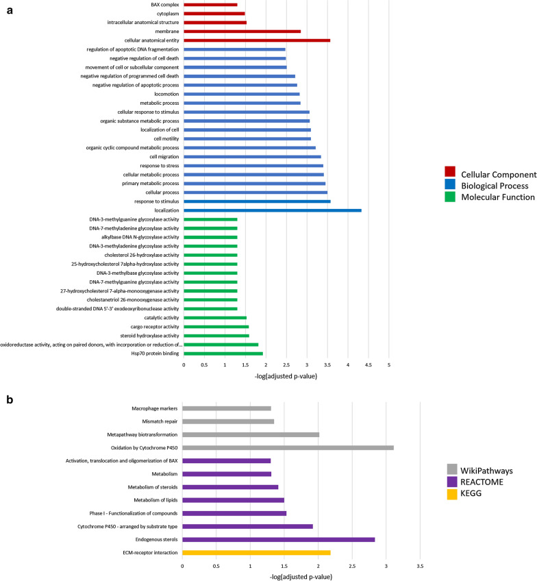
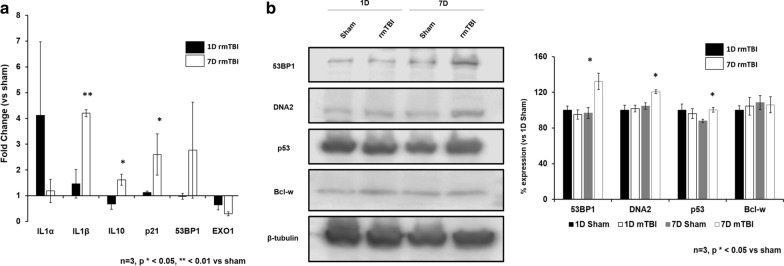
Mild traumatic brain injury (mTBI) results in broad neurological symptoms and an increased risk of being diagnosed with a neurodegenerative disease later in life. While the immediate oxidative stress response and post-mortem pathology of the injured brain has been well studied, it remains unclear how early pathogenic changes may drive persistent symptoms and confer susceptibility to neurodegeneration. In this study we have used a mouse model of repeated mTBI (rmTBI) to identify early gene expression changes at 24 h or 7 days post-injury (7 dpi). At 24 h post-injury, gene expression of rmTBI mice shows activation of the DNA damage response (DDR) towards double strand DNA breaks, altered calcium and cell-cell signalling, and inhibition of cell death pathways. By 7 dpi, rmTBI mice had a gene expression signature consistent with induction of cellular senescence, activation of neurodegenerative processes, and inhibition of the DDR. At both timepoints gliosis, microgliosis, and axonal damage were evident in the absence of any gross lesion, and by 7 dpi rmTBI also mice had elevated levels of IL1β, p21, 53BP1, DNA2, and p53, supportive of DNA damage-induced cellular senescence. These gene expression changes reflect establishment of processes usually linked to brain aging and suggests that cellular senescence occurs early and most likely prior to the accumulation of toxic proteins. These molecular changes were accompanied by spatial learning and memory deficits in the Morris water maze. To conclude, we have identified DNA damage-induced cellular senescence as a repercussion of repeated mild traumatic brain injury which correlates with cognitive impairment. Pathways involved in senescence may represent viable treatment targets of post-concussive syndrome. Senescence has been proposed to promote neurodegeneration and appears as an effective target to prevent long-term complications of mTBI, such as chronic traumatic encephalopathy and other related neurodegenerative pathologies.

Keywords: Ageing; Concussion; DNA damage response; Neurodegeneration; Neuroinflammation; Senescence; Traumatic brain injury.


Conflict of interest statement

The authors declare that they have no competing interests.

Figures

Fig. 1
Fig. 1
The duration of loss of righting reflex in rmTBI was significantly longer than in sham-treated mice after each impact (p < 0.001 main effect of injury status, repeated measures ANOVA), and decreased significantly with increased impact number (p < 0.001, interaction between injury status and impact number, repeated measures ANOVA). Righting reflex was under 15 min for all mice, supportive of a mild injury
Fig. 2
Fig. 2
rmTBI mice show evidence of spatial learning and memory impairment at 1 week post-injury. a rmTBI mice swam an average of 1.8 cm/s faster than sham mice. b Across three training days rmTBI mice had a significantly increased latency to find the platform compared to shams (p < 0.001, main effect of injury status, repeated measures ANOVA). c In the probe test rmTBI mice spent less time in the platform zone compared to shams (p = 0.55, Mann–Whitney U Test). Analysis of search strategies with Rtrack revealed no significant differences one training day one, however rmTBI mice used significantly different search strategies by training days two (χ2 = 6.2, df = 2, p = 0.05) and three (χ2 = 33.9, df = 2, p < 0.0001) with rmTBI mice utilizing less allocentric strategies than shams by the end of training
Fig. 3
Fig. 3
Gross morphology of the mouse brain following Sham (a) and rmTBI (b) procedures. H&E staining of the rmTBI brain (c) reveals no visible lesion induced by the injury model, indicating a mild injury has been elicited
Fig. 4
Fig. 4
rmTBI brains have increased expression of GFAP at 1 day (b) and 7 days (c) post-injury, compared to shams (a) in the impact region of the isocortex, indicating gliosis. Microglial activation, shown with Iba1 staining, can be seen in rmTBI brains at both 1 day (e) and 7 days (f) post-injury compared to shams (d). At 7 days post-injury rmTBI mice showed evidence of axonal damage by the presence of APP positive spheroids in the subcortical white matter (h), which was not present in shams (i, low power view in g). Scale bars: 0.3 mm in af, 0.1 mm in g, 0.05 mm in h, 0.08 mm in i
Fig. 5
Fig. 5
Lamin A/C expression on the nuclear membrane of glial cells was reduced at 1 day post injury in the cortex (b) and in neurons of the dentate (e), and this loss was exacerbated by 1 week post-injury in the cortex (c) and dentate (f) compared to shams (a, d). Arrows in b and c point to glial cells that have lost lamin expression. Scale bar: 150 μm in ac, 300 μm in d, 120 μm in e, f
Fig. 6
Fig. 6
Overview of significantly (p ≤ 0.05) differentially expressed genes between rmTBI and sham mice at 24 h post-injury
Fig. 7
Fig. 7
Significantly (p ≤ 0.05) upregulated genes in rmTBI mice compared to shams at 24 h post-injury. Data are expressed as normalized RNA count number for shams and rmTBI. The median value is represented by the horizontal line within the box, defined by the first and third quartiles, and tails represent 1.5 × the interquartile range
Fig. 8
Fig. 8
Gene set enrichment analysis of genes significantly (p ≤ 0.05) upregulated 24 h post-injury. a Gene ontology (GO) functions significantly enriched include DNA damage response pathways and b pathways significantly enriched include the DNA damage response and cell-cycle checkpoints
Fig. 9
Fig. 9
Significantly (p ≤ 0.05) downregulated genes in rmTBI mice compared to shams at 24 h post-injury. Data are expressed as normalized RNA count number for shams and rmTBI. The median value is represented by the horizontal line within the box, defined by the first and third quartiles, and tails represent 1.5 × the interquartile range
Fig. 10
Fig. 10
Gene set enrichment analysis of genes significantly (p ≤ 0.05) upregulated 24 h post-injury. a Gene ontology (GO) functions and b pathway analysis suggest inhibition of cell death pathways and altered metabolic function
Fig. 11
Fig. 11
Overview of significantly (p ≤ 0.05) differentially expressed genes between rmTBI and sham mice at 7d post-injury
Fig. 12
Fig. 12
Significantly (p ≤ 0.05) upregulated genes in rmTBI mice compared to shams at 7d post-injury. Data are expressed as normalized RNA count number for shams and rmTBI. The median value is represented by the horizontal line within the box, defined by the first and third quartiles, and tails represent 1.5 × the interquartile range
Fig. 13
Fig. 13
Significantly (p ≤ 0.05) downregulated genes in rmTBI mice compared to shams at 7d post-injury. Data are expressed as normalized RNA count number for shams and rmTBI. The median value is represented by the horizontal line within the box, defined by the first and third quartiles, and tails represent 1.5 × the interquartile range
Fig. 14
Fig. 14
Gene ontology (a) and pathway (b) enrichment analyses of genes significantly (p ≤ 0.05) upregulated 7d post-injury
Fig. 15
Fig. 15
Gene ontology (a) and pathway (b) enrichment analyses of genes significantly (p ≤ 0.05) downregulated 7d post-injury
Fig. 16
Fig. 16
a qPCR and b Western Blot validation. Statistical analysis done in R using unpaired student t-test vs sham

Similar articles

Cited by

References

    1. Alemi M, Gaiteiro C, Ribeiro CA, Santos LM, Gomes JR, Oliveira SM, et al. Transthyretin participates in beta-amyloid transport from the brain to the liver–involvement of the low-density lipoprotein receptor-related protein 1? Sci Rep. 2016;6:20164. doi: 10.1038/srep20164. - DOI - PMC - PubMed
    1. Armistead-Jehle P, Soble JR, Cooper DB, Belanger HG. Unique aspects of traumatic brain injury in military and veteran populations. Phys Med Rehabil Clin N Am. 2017;28(2):323–337. doi: 10.1016/j.pmr.2016.12.008. - DOI - PubMed
    1. Arun P, Rossetti F, Wilder DM, Sajja S, Van Albert SA, Wang Y, et al. Blast exposure leads to accelerated cellular senescence in the rat brain. Front Neurol. 2020;11:438. doi: 10.3389/fneur.2020.00438. - DOI - PMC - PubMed
    1. Atamna H, Nguyen A, Schultz C, Boyle K, Newberry J, Kato H, et al. Methylene blue delays cellular senescence and enhances key mitochondrial biochemical pathways. FASEB J. 2008;22(3):703. doi: 10.1096/fj.07-9610com. - DOI - PubMed
    1. Bakkenist CJ, Drissi R, Wu J, Kastan MB, Dome JS. Disappearance of the telomere dysfunction-induced stress response in fully senescent cells. Cancer Res. 2004;64(11):3748. doi: 10.1158/0008-5472.CAN-04-0453. - DOI - PubMed

Publication types

Grants and funding