Review
. 2021 May;147(5):1531-1547.
doi: 10.1016/j.jaci.2021.03.015.

Roles of innate lymphoid cells (ILCs) in allergic diseases: The 10-year anniversary for ILC2s

Affiliations
Review

Roles of innate lymphoid cells (ILCs) in allergic diseases: The 10-year anniversary for ILC2s

Kathleen R Bartemes et al. J Allergy Clin Immunol. 2021 May.

Abstract

In the 12 years since the discovery of innate lymphoid cells (ILCs), our knowledge of their immunobiology has expanded rapidly. Group 2 ILCs (ILC2s) respond rapidly to allergen exposure and environmental insults in mucosal organs, producing type 2 cytokines. Early studies showed that epithelium-derived cytokines activate ILC2s, resulting in eosinophilia, mucus hypersecretion, and remodeling of mucosal tissues. We now know that ILC2s are regulated by other cytokines, eicosanoids, and neuropeptides as well, and interact with both immune and stromal cells. Furthermore, ILC2s exhibit plasticity by adjusting their functions depending on their tissue environment and may consist of several heterogeneous subpopulations. Clinical studies show that ILC2s are involved in asthma, allergic rhinitis, chronic rhinosinusitis, food allergy, and eosinophilic esophagitis. However, much remains unknown about the immunologic mechanisms involved. Beneficial functions of ILCs in maintenance or restoration of tissue well-being and human health also need to be clarified. As our understanding of the crucial functions ILCs play in both homeostasis and disease pathology expands, we are poised to make tremendous strides in diagnostic and therapeutic options for patients with allergic diseases. This review summarizes discoveries in immunobiology of ILCs and their roles in allergic diseases in the past 5 years, discusses controversies and gaps in our knowledge, and suggests future research directions.

Keywords: Innate lymphoid cells; allergic rhinitis; asthma; atopic dermatitis; chronic rhinosinusitis; eosinophilic esophagitis; group 2 innate lymphoid cells.


Figures

Figure 1.
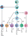
Figure 1.. Development of ILCs.
ILCs begin as common lymphoid progenitors (CLP) which differentiate into common innate lymphoid progenitors (CILP) under the control of Id2. CILPs can differentiate into common helper innate lymphoid progenitors (CHILP) or into NK cell precursors (NKP). NKPs further differentiate into NK cells while CHILPs can follow one of three programs. First, lymphoid tissue inducer progenitors (LTiP) arise from CHILPs and can give rise to lymphoid tissue inducer cells (LTi). Second, CHILPs under the influence of Id3 differentiate into regulatory innate lymphoid cells (ILCreg). Third, CHILPs give rise to innate lymphoid cell precursors (ILCP). ILCPs differentiate into ILC1, ILC2 or ILC3 subsets regulated by the lineage-specific transcription factors T-bet, GATA3/RORα, or RORγt/AHR, respectively. Note that differentiated ILC subsets maintain some degree of plasticity as represented in Figure 3 for ILC2s. AHR, aryl hydrocarbon receptor; Areg, amphiregulin; EOMES, Eomesodermin; GATA3, GATA Binding Protein 3; GM-CSF, granulocyte macrophage colony-stimulating factor; Id2, inhibitor of DNA binding 2; Id3, inhibitor of DNA binding 3; IFN-γ, interferon-γ; IL, interleukin; ILC, innate lymphoid cell; NK, natural killer; RORα, RAR-related orphan receptor a; RORγt, RAR-related orphan receptor γt; T-bet, T-box transcription factor; TGF-β, transforming growth factor–β; TNF-α, tumor necrosis factor-a; TSLP, thymic stromal lymphopoietin
Figure 2.
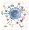
Figure 2.. Factors that activate and inhibit ILC2s and their potential cellular sources with focus on allergic immune responses. Activation.
Activation of ILC2s by cytokines, eicosanoids and neuropeptides generated by epithelial cells, keratinocytes, macrophages, dendritic cells, Tuft cells, brush cells, and neurons in allergic diseases has been demonstrated in both mice and humans. Inhibition. Epithelial cells, NK cells, DCs, Tregs, ILCregs, neurons and neutrophils inhibit ILC2s both by direct interaction and by generation of cytokines, eicosanoids and neuropeptides. CD62P, P-selectin; CGRP, calcitonin gene-related peptide; cysLT, cysteinyl leukotriene; DC, dendritic cell; DR3, death receptor 3; FFA, free fatty acid; G-CSF, granulocyte colony-stimulating factor; IFN-α, interferon-α IFN-β, interferon-β; IFN-γ, interferon-γ; IL, interleukin; LTC4, leukotriene C4; LTD4, leukotriene D4; NK, natural killer; NMU, neuromedin U; PGD2, prostaglandin D2; PGE2, prostaglandin E2; PGI2, prostaglandin I2; PMN MDSC, polymorphonuclear myeloid-derived suppressor cell; PSGL-1, P-selectin glycoprotein ligand-1; SCF, stem cell factor; TGF-β, transforming growth factor–β; TL1A, tumor necrosis factor-like cytokine 1A; Treg, regulatory T; TSLP, thymic stromal lymphopoietin; VIP, vasoactive intestinal peptide
Figure 3.
Figure 3.. Plasticity of ILC2s.
ILC2s can be directed by cytokines to change their abilities to produce certain sets of cytokines. While ILC2s regularly produce type 2 cytokines and amphiregulin, they can be re-programmed to produce IFN-γ (i.e., ILC1-like) when stimulated with IL-12 and IL-1β. Importantly, ILC1-like ILC2s retain the ability to produce IL-13, and this transition can be reversed with IL-4. ILC2s can be induced to produce IL-10 (i.e., IL-10+ ILC2s) when cultured with IL-4 or RA in the presence of a cocktail of IL-33, IL-2 and IL-17. ILC2s can produce IL-17 when stimulated with IL-1β, IL-6, IL-23 and TGF-β (i.e., ILC3-like). Note that our knowledge of ILC2 plasticity is in an early stage, and further studies are necessary to identify the optimal conditions to drive transition of ILC2s from one subset to another in vivo and in vitro and to uncover the mechanisms involved. Areg, amphiregulin; IFN-γ, interferon-γ; IL, interleukin; RA, retinoic acid; TGF-β, transforming growth factor–β

References

    1. Pulendran B, Artis D. New paradigms in type 2 immunity. Science. 2012;337:431–5. - PMC - PubMed
    1. Boonpiyathad T, Sozener ZC, Satitsuksanoa P, Akdis CA. Immunologic mechanisms in asthma. Semin Immunol. 2019;46:101333. - PubMed
    1. Fallon PG, Ballantyne SJ, Mangan NE, Barlow JL, Dasvarma A, Hewett DR, et al. Identification of an interleukin (IL)-25-dependent cell population that provides IL-4, IL-5, and IL-13 at the onset of helminth expulsion. J Exp Med. 2006;203:1105–16. - PMC - PubMed
    1. Fort MM, Cheung J, Yen D, Li J, Zurawski SM, Lo S, et al. IL-25 induces IL-4, IL-5, and IL-13 and Th2-associated pathologies in vivo. Immunity. 2001; 15:985–95. - PubMed
    1. Moro K, Yamada T, Tanabe M, Takeuchi T, Ikawa T, Kawamoto H, et al. Innate production of T(H)2 cytokines by adipose tissue-associated c-Kit(+)Sca-1(+) lymphoid cells. Nature. 2010;463:540–4. - PubMed

Publication types