

Association of toe-extension movement pattern magnitude and variability during three functional tasks with diabetic foot complications
- PMID: 33965738
- PMCID: PMC8283772
- DOI: 10.1016/j.clinbiomech.2021.105371
Association of toe-extension movement pattern magnitude and variability during three functional tasks with diabetic foot complications
Abstract
Background: A toe-extension movement pattern may contribute to metatarsophalangeal joint deformity and ulceration in people with diabetes. We sought to quantify the relationship between toe extension magnitude and variability during three functional tasks (ankle range of motion, sit to stand, walking) with metatarsophalangeal joint deformity, and identify potential mechanisms associated with a toe-extension movement pattern.
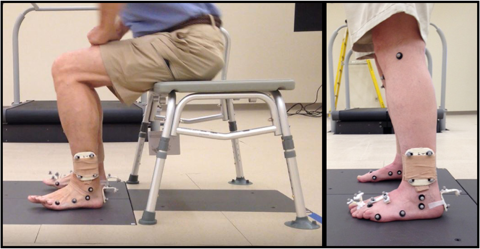
Methods: Individuals with diabetes and peripheral neuropathy were included (n = 60). Metatarsophalangeal joint deformity was assessed using computed tomography (CT). Toe-extension movement was quantified using 3-dimensional motion capture. Linear regression was used to investigate the role of toe-extension movement pattern on metatarsophalangeal joint deformity. Regression analysis was used to identify mechanisms (neuropathy severity, foot intrinsic muscle deterioration ratio, ankle dorsiflexion range of motion) contributing to toe-extension movement pattern.
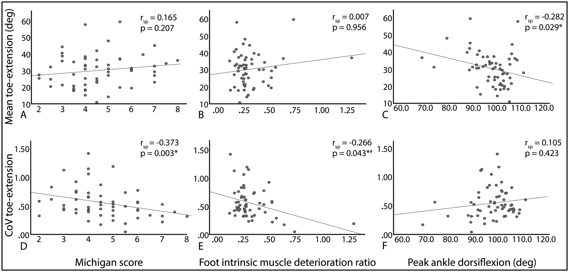
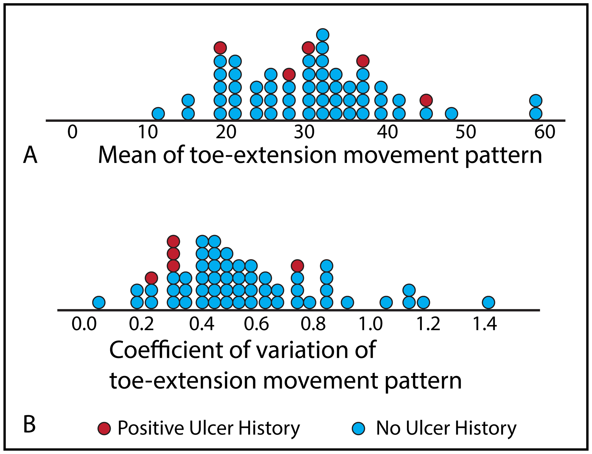
Findings: Toe extension with each functional task as well as the mean and coefficient of variation across all tasks were significantly related to metatarsophalangeal joint deformity (range of correlation coefficients = (-0.386, 0.692), p ≤ 0.001). Ankle dorsiflexion range of motion was associated with mean toe extension across all tasks (rsp = -0.282, p = 0.029). Neuropathy severity and foot intrinsic muscle deterioration ratio were associated with toe extension variability (rsp = -0.373, p = 0.003 and rsp = -0.266, p = 0.043; respectively).
Interpretation: Greater magnitude and lower variability of a toe-extension movement pattern was found to be associated with metatarsophalangeal joint deformity. These findings may support clinical assessment and treatment of movement across more than one task.
Keywords: Ankle; Biomechanics; Foot; Gait; Neuropathy; Rehabilitation.
Copyright © 2021. Published by Elsevier Ltd.
Figures
References
-
- American Diabetes Association, 2003. Peripheral arterial disease in people with diabetes. Diabetes Care 26, 3333–3341. - PubMed
-
- Armstrong D, Lavery L, Vela S, Quebedeaux T, Fleischli J, 1998. Choosing a practical screening instrument to identify patients at risk for diabetic foot ulceration. Arch. Intern. Med 158, 289–292. - PubMed
-
- Armstrong DG, Boulton AJM, Bus SA, 2017. Diabetic foot ulcers and their recurrence. N. Engl. J. Med 376, 2367–2375. - PubMed
-
- Brooks B, Dean R, Patel S, Wu B, Molyneaux L, Yue DK, 2001. TBI or not TBI: That is the question. Is it better to measure toe pressure than ankle pressure in diabetic patients? Diabet. Med 18, 528–532. - PubMed
-
- Cavanagh PR, Perry JE, Ulbrecht JS, Derr JA, Pammer SE, 1998. Neuropathic diabetic patients do not have reduced variability of plantar loading during gait. Gait Posture 7, 191–199. - PubMed
Publication types
MeSH terms
Grants and funding
LinkOut - more resources
Full Text Sources
Other Literature Sources
Medical
