

doi: 10.1038/s41392-021-00574-8.
SARS-CoV-2 promote autophagy to suppress type I interferon response
Affiliations
- PMID: 33966045
- PMCID: PMC8105701
- DOI: 10.1038/s41392-021-00574-8
Item in Clipboard
SARS-CoV-2 promote autophagy to suppress type I interferon response
Signal Transduct Target Ther.
.
No abstract available
Conflict of interest statement
The authors declare no competing interests.
Figures
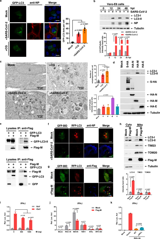
SARS-CoV-2 promote autophagy to suppress type I interferon response. a GFP-LC3 dot formation in Vero-E6 cells transiently transfected with GFP-LC3 and either left uninfected (Mock) or infected with SARS-CoV-2 (MOI of 0.05) for 48 h or treated with CQ for 4 h. Scale bar, 10 µm. b Vero-E6 were uninfected (−) or infected (+) with SARS-CoV-2. Lysates were evaluated by western blotting (WB). c EM analysis of Vero-E6 cells that were stimulated with CQ for 4 h, or infected with SARS-CoV-2 (MOI of 0.05) for 24 h. Scale bar, 2 µm. d Huh7.0 cells were transfected with vector or S-HA, HA-M, HA-N, or HA-E plasmid. Lysates were analysed by immunoblotting. e Interaction between Flag-M and GFP-LC3 in HEK293T cells. f Huh7.0 cells were transfected with the indicated plasmids and infected with SARS-CoV-2 and analysed for the co-localization of BID-GFP and RFP-LC3. g Huh7.0 cells were transfected with the indicated plasmids and analysed for the co-localization of BID-GFP and RFP-LC3. h Huh7.0 cells were transfected with Flag-M plasmid, and mitochondrial fractions were isolated via ultracentrifugation. Cytoplasm (Cyto) and mitochondria (Mito) were analysed by immunoblotting. i HEK293T cells were transfected with the indicated plasmids and infected with Sendai virus for 8 h before the reporter assay was conducted. j HEK293T cells were transfected with the indicated plasmids, and a reporter assay was conducted after transfection. k HEK293T cells were transfected with the indicated plasmids, and a reporter assay was conducted after transfection. Three independent experiments with three technical repetitions were performed. Data are expressed as mean ± SEM (error bars). Statistical analyses used Student’s t test. P < 0.05 was considered statistically significant
Similar articles
-
SARS-CoV-2 genomic surveillance identifies naturally occurring truncation of ORF7a that limits immune suppression.Cell Rep. 2021 Jun 1;35(9):109197. doi: 10.1016/j.celrep.2021.109197. Epub 2021 May 14. Cell Rep. 2021. PMID: 34043946 Free PMC article.
-
A systemic and molecular study of subcellular localization of SARS-CoV-2 proteins.Signal Transduct Target Ther. 2020 Nov 17;5(1):269. doi: 10.1038/s41392-020-00372-8. Signal Transduct Target Ther. 2020. PMID: 33203855 Free PMC article. No abstract available.
-
Systematic functional analysis of SARS-CoV-2 proteins uncovers viral innate immune antagonists and remaining vulnerabilities.Cell Rep. 2021 May 18;35(7):109126. doi: 10.1016/j.celrep.2021.109126. Epub 2021 Apr 27. Cell Rep. 2021. PMID: 33974846 Free PMC article.
-
SARS-CoV-2 infection and the antiviral innate immune response.J Mol Cell Biol. 2020 Nov 26;12(12):963-967. doi: 10.1093/jmcb/mjaa071. J Mol Cell Biol. 2020. PMID: 33377937 Free PMC article. Review. No abstract available.
-
Type I interferon pathway genetic variants in severe COVID-19.Virus Res. 2024 Apr;342:199339. doi: 10.1016/j.virusres.2024.199339. Epub 2024 Feb 22. Virus Res. 2024. PMID: 38354910 Free PMC article. Review.
Cited by
-
PRRSV utilizes MALT1-regulated autophagy flux to switch virus spread and reserve.Autophagy. 2024 Dec;20(12):2697-2718. doi: 10.1080/15548627.2024.2386195. Epub 2024 Aug 6. Autophagy. 2024. PMID: 39081059 Free PMC article.
-
The role of autophagy in viral infections.J Biomed Sci. 2023 Jan 18;30(1):5. doi: 10.1186/s12929-023-00899-2. J Biomed Sci. 2023. PMID: 36653801 Free PMC article. Review.
-
Autophagy in Inflammatory Response against SARS-CoV-2.Int J Mol Sci. 2023 Mar 3;24(5):4928. doi: 10.3390/ijms24054928. Int J Mol Sci. 2023. PMID: 36902354 Free PMC article. Review.
-
A Comparative Transcriptomic Analysis Reveals That HSP90AB1 Is Involved in the Immune and Inflammatory Responses to Porcine Deltacoronavirus Infection.Int J Mol Sci. 2022 Mar 18;23(6):3280. doi: 10.3390/ijms23063280. Int J Mol Sci. 2022. PMID: 35328701 Free PMC article.
-
The intestinal microbial metabolite nicotinamide n-oxide prevents herpes simplex encephalitis via activating mitophagy in microglia.Gut Microbes. 2022 Jan-Dec;14(1):2096989. doi: 10.1080/19490976.2022.2096989. Gut Microbes. 2022. PMID: 35793266 Free PMC article.
References
Publication types
MeSH terms
Substances
Grants and funding
- 2662020PY002/Huazhong Agricultural University (HZAU)
- 81825015/National Natural Science Foundation of China (National Science Foundation of China)
- 81871650/National Natural Science Foundation of China (National Science Foundation of China)
- 31630086/National Natural Science Foundation of China (National Science Foundation of China)
- 2017CFA022/Natural Science Foundation of Hubei Province (Hubei Provincial Natural Science Foundation)
LinkOut - more resources
Full Text Sources
Other Literature Sources
Medical
Molecular Biology Databases
Miscellaneous
