Review
. 2021 May 10;7(5):1725-1741.
doi: 10.1021/acsbiomaterials.0c01132. Epub 2020 Oct 14.

Nano- and Microformulations to Advance Therapies for Visceral Leishmaniasis

Affiliations
Review

Nano- and Microformulations to Advance Therapies for Visceral Leishmaniasis

Devika M Varma et al. ACS Biomater Sci Eng. .

Abstract

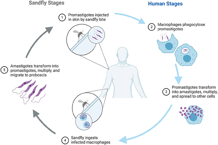
Visceral leishmaniasis (VL) is a deadly, vector-borne, neglected tropical disease endemic to arid parts of the world and is caused by a protozoan parasite of the genus Leishmania. Chemotherapy is the primary treatment for this systemic disease, and multiple potent therapies exist against this intracellular parasite. However, several factors, such as systemic toxicity, high costs, arduous treatment regimen, and rising drug resistance, are barriers for effective therapy against VL. Material-based platforms have the potential to revolutionize chemotherapy for leishmaniasis by imparting a better pharmacokinetic profile and creating patient-friendly routes of administration, while also lowering the risk for drug resistance. This review highlights promising drug delivery strategies and novel therapies that have been evaluated in preclinical models, demonstrating the potential to advance chemotherapy for VL.

Keywords: Leishmania donovani; Leishmania infantum; emulsions; liposomes; micelles; nanoparticles; polymeric particles; polymersomes; visceral leishmaniasis.


Conflict of interest statement

The authors declare the following competing financial interest(s): Drs. Ainslie and Bachelder serve on the advisory board for IMMvention Therapeutix, Inc. Although a financial conflict of interest was identified for management based on the overall scope of the project and its potential benefit to IMMvention Therapeutix, Inc., the research findings included in this publication may not necessarily relate to the interests of IMMvention Therapeutix, Inc.

Figures

Figure 1.
Figure 1.
Schematic showing dual life stages of Leishmania. Sandflies carrying the promastigote forms of the parasite infect humans through a bite. Inside the human body, promastigotes phagocytosed by macrophages transform into amastigotes and multiply. The parasite lifecycle continues, as another sandfly ingests infected host cells upon biting an infected mammalian host, where the amastigotes transform into promastigotes in the midgut.
Figure 2.
Figure 2.
Schematic showing in vitro and in vivo evaluations of an experimental drug formulation. (A) Parasites used in experiments are initially isolated from spleens of infected hamster, where splenic amastigotes are differentiated into promastigotes. (B) In vitro intracellular evaluation. Typically, mouse-derived macrophages are infected with promastigotes, treated with drug formulation, and enumerated via Giemsa stain. (C) Anti-parasitic effects can be evaluated directly against promastigotes and axenic amastigotes. Spectrometric measurements can be used to evaluate parasite viability by either staining the parasite or using genetically modified, fluorescent or luminescent strains of Leishmania. (D) In vivo, mice are infected with promastigotes and treated once infection is established. The parasite load after treatment is determined using Giemsa-stained smears from the liver, spleen, and bone marrow. The parasitic burden is often expressed as Leishman–Donovan units (LDU).
Figure 3.
Figure 3.
Schematic representing two types of primary emulsions. (A) The oil-in-water (o/w) emulsion (left) shows a single layer of phospholipids with hydrophobic tails facing inward, creating a lipophilic environment for the oil and hydrophobic drug in the center. The water-in-oil emulsion droplet (right) shows the lipid tails facing outward. The hydrophilic heads surround the water with hydrophilic drug in the center. (B) Emulsion method. A mixture of amphiphile, drug, and buffer is typically sonicated or homogenized to create an even dispersion of droplets throughout the emulsion. In some cases, the pH is adjusted to stabilize the emulsion. Finally, the emulsion is often filtered through a membrane filter to ensure homogeneity of the emulsion size.
Figure 4.
Figure 4.
Das et al. formulated ursolic acid (UA) in a nanostructured lipid carrier with a surface coating of N-octyl-chitosan (UA-NLC). (A) Schematic representing a drug-loaded lipid matrix surrounded by amphiphilic polymer. (B) TEM image of UA-NLC. Scale bar =100 nm. (C) Percent suppression of splenic parasitic burden in L. donovani-infected BALB/c mice post treatment with UA-NLC. Wild-type, drug-resistant (SSG-R and PMM-R), and GE1 field-type strains of L. donovani were used to infect mice. Adapted with permission from ref . Copyright 2017 Elsevier B.V.
Figure 5.
Figure 5.
Micellar formulations. (A) Schematic of a lipid micelle carrying a hydrophobic drug in an oily core. The structure of a single phospholipid is shown. (B) A polymeric micelle, with hydrophobic and hydrophilic sections of monomer shown to the right. (C) A cryo-TEM image of polymeric micelles fabricated by the emulsion solvent evaporation method. Scale bar = 200 nm. (D) Micellar formation via emulsion solvent evaporation. Drug and micellar components are combined in an organic solution. The mixture is sonicated or homogenized to create an emulsion. In a rotary evaporator, the organic solution evaporates from the emulsion droplets, transforming them to micelles. Reproduced with permission from ref . Copyright 2005 Elsevier B.V.
Figure 6.
Figure 6.
Singh et al. encapsulated amphotericin B (AmpB) in chitosan-coated Pluronic F127 micelles (AmB-Ch-PF-M) in an attempt to target macrophages (Cs-PF-AmB-M). Shown here are AmB concentrations in the (A) liver and (B) spleen in infected hamsters treated with Amp B formulations, liposomal Amp B (LAmpB), AmB-Ch_PF-M, or uncoated micelles (AmB-PF-M). Reproduced with permission from ref . Copyright 2017 Elsevier B.V.
Figure 7.
Figure 7.
Liposomal formulations. (A) Schematic of a liposome. The phospholipid bilayer encircles the aqueous core of the liposome, allowing transport of hydrophobic and hydrophilic drugs. The structure of a single phospholipid is shown. (B) A cryo-TEM image of blank liposomes fabricated by thin lipid film hydration. Scale bar = 100 nm. (C) Liposome formation via thin lipid film hydration. Organic solvents are evaporated from the solution with lipid components until a dried lipid film is left. The film is hydrated with aqueous solvent, typically with a drug. After hydration, heterogeneous liposomes form in solution and encapsulate the drug, passively or via remote loading. Sonication or extrusion is applied to the solution to obtain homogeneous liposomes of the desired size. Reproduced with permission from ref . Copyright 2017 Springer.
Figure 8.
Figure 8.
Gupta et al. formulated liposome-encapsulated pentalinonsterol (PEN). Treatment in L. donovani-infected mice triggered a host-protective Th1 immune response. (A) Structure of synthesized PEN. (B) Hepatic granuloma formation in infected mice. Mice treated with liposomal PEN showed better-formed granulomas with less parasites than those treated with empty liposomes or PBS (400×). Liposomal PEN treatment effectively decreases parasitic loads in the (C) liver, (D) spleen, and (E) bone marrow of infected mice. ***p < 0.001. Reproduced with permission from ref . Copyright 2017 American Chemical Society.
Figure 9.
Figure 9.
Polymeric matrix nanoparticle formulation. (A) Schematic of a polymeric NP that can carry hydrophobic or hydrophilic drugs. (B) An SEM image of nanoprecipitated NPs coated with Dextran 40. Scale bar = 500 nm. (C) Method of nanoprecipitation. The polymer and drug in an organic phase are dropped into an aqueous phase containing a particle stabilizer and surfactant in order to create an emulsion of organic and aqueous solvents. With magnetic stirring, the organic solvent evaporates, allowing the particles to harden. Finally, the particles are isolated by centrifugation. Reproduced with permission from ref . Copyright 2010 Springer.
Figure 10.
Figure 10.
Formulation AR-12 in acetalated dextran (Ac-DEX) microparticles (AR-12/MPs). (A) SEM image of AR-12/MPs. (B) Clearance of L. donovani promastigotes in vitro after 72 h with different treatments. Leishmania–Donovan units (LDU) in the (C) liver and (D) spleen of L. donovani-infected BALB/c mice treated 28 days post infection with sodium stibogluconate (SSG), PBS, AR-12/MPs, and amphotericin B (AmpB). Mice treated with AR-12/MPs have significantly lower liver and spleen LDUs than those treated with PBS. *p < 0.05, **p < 0.01, ***p < 0.005 with respect to AR-12/MPs + AmpB 1.0 mg/kg. Reproduced with permission from ref . Copyright 2014 American Chemical Society.
Figure 11.
Figure 11.
Gupta et al. synthesized polymersome with glycol chitosan-stearic acid copolymers (GC-SA) carrying lipoteichoic acid (LTA) functionalized AmB (LTA-AmB-L-Psome). (A) Schematic of the formulated polymersome. (B) TEM image of AmB polymersomes with LTA functionalization. Scale bar = 200 nm. (C) Comparison of parasitic inhibition by different formulations (AmB-L-Psome, AmB-loaded polymersome; L-Psome, polymersome; LTA-L-Psome, LTA carrying polymersome) tested in vivo in L. donovani-infected hamsters. *p < 0.05, **p < 0.01, ***p < 0.001. Adapted with permission from ref . Copyright 2014 American Chemical Society.

Similar articles

Cited by

References

    1. Burza S; Croft SL; Boelaert M Leishmaniasis. Lancet 2018, 392 (10151), 951–970. - PubMed
    1. Olivier M; Badaró R; Medrano FJ; Moreno J The pathogenesis of Leishmania/HIV co-infection: cellular and immunological mechanisms. Ann. Trop. Med. Parasitol 2003, 97 (Suppl 1), 79–98. - PubMed
    1. Jarvis JN; Lockwood DN Clinical aspects of visceral leishmaniasis in HIV infection. Curr. Opin. Infect. Dis 2013, 26 (1), 1–9. - PubMed
    1. Zijlstra EE The immunology of post-kala-aza dermal leishmaniasis (PKDL). Parasites Vectors 2016, 9, 464. - PMC - PubMed
    1. Ghosh M; Roy K; Roy S Immunomodulatory effects of antileishmanial drugs. J. Antimicrob. Chemother 2013, 68 (12), 2834–8. - PubMed

Publication types

LinkOut - more resources