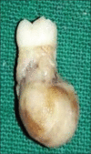
Cementoblastoma of a primary molar: A rare pediatric occurrence
- PMID: 33967495
- PMCID: PMC8083404
- DOI: 10.4103/jomfp.JOMFP_307_19
Cementoblastoma of a primary molar: A rare pediatric occurrence
Abstract
Cementoblastoma is a relatively uncommon, benign odontogenic mesenchymal tumor that is associated with and attached to the roots of teeth. It is considered to be the only true neoplasm of cemental origin. Its prevalence has been reported to vary from 0.69% to 8% of all odontogenic tumors. The tumor is frequently seen in the second and third decades of life and affects the molar and premolar regions of the mandible predominantly. We herein describe a case of cementoblastoma occurring in association with primary teeth in a 5-year-old male patient with a brief review of literature. Only 20 cases of cementoblastoma involving primary teeth were found after the English literature search, the current case being the 21st. Moreover, the deciduous teeth-associated cementoblastomas (14 out of 20) show a distinct predilection for the right side of the face. The current case is the seventh one to involve the left side.
Keywords: Cementoblastoma; cementum; odontogenic tumor; primary molar; tooth root.
Copyright: © 2021 Journal of Oral and Maxillofacial Pathology.
Conflict of interest statement
There are no conflicts of interest.
Figures


Similar articles
-
Cementoblastoma Solely Involving Maxillary Primary Teeth--A Rare Presentation.J Clin Pediatr Dent. 2016;40(2):147-51. doi: 10.17796/1053-4628-40.2.147. J Clin Pediatr Dent. 2016. PMID: 26950817
-
Benign cementoblastoma involving left deciduous first molar: A case report and review of literature.J Oral Maxillofac Pathol. 2019 Sep-Dec;23(3):422-428. doi: 10.4103/jomfp.JOMFP_193_19. J Oral Maxillofac Pathol. 2019. PMID: 31942125 Free PMC article.
-
Benign cementoblastoma involving multiple maxillary teeth: report of a case with a review of the literature.Oral Surg Oral Med Oral Pathol Oral Radiol Endod. 2004 Jan;97(1):53-8. doi: 10.1016/j.tripleo.2003.08.012. Oral Surg Oral Med Oral Pathol Oral Radiol Endod. 2004. PMID: 14716256 Review.
-
A Rare Case of Cementoblastoma of the Second Right Maxillary Premolar in a 30-Year-Old Man.Cureus. 2024 Nov 15;16(11):e73737. doi: 10.7759/cureus.73737. eCollection 2024 Nov. Cureus. 2024. PMID: 39677262 Free PMC article.
-
Benign cementoblastoma. Case report.Aust Dent J. 1996 Feb;41(1):9-11. doi: 10.1111/j.1834-7819.1996.tb05647.x. Aust Dent J. 1996. PMID: 8639125 Review.
References
-
- Chaput A, Marc A. A case of cementoma localized on a temporary molar. SSO Schweiz Monatsschr Zahnheilkd. 1965;75:48–52. - PubMed
-
- Vilasco J, Mazère J, Douesnard JC, Loubière R. A case of cementoblastoma. Rev Stomatol Chir Maxillofac. 1969;70:329–32. - PubMed
-
- Zachariades N, Skordalaki A, Papanicolaou S, Androulakakis E, Bournias M. Cementoblastoma: Review of the literatureand report of a case in a seven-year-old girl. Br J Oral Maxillofac Surg. 1985;23:456–61. - PubMed
-
- Herzog S. Benign cementoblastoma associated with the primary dentition. J Oral Med. 1987;42:106–8.
Publication types
LinkOut - more resources
Full Text Sources
