

Rare histologic presentation of pleomorphic adenoma: A diagnostic dilemma
- PMID: 33967498
- PMCID: PMC8083414
- DOI: 10.4103/jomfp.JOMFP_62_20
Rare histologic presentation of pleomorphic adenoma: A diagnostic dilemma
Abstract
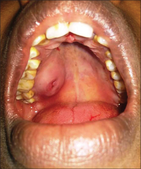
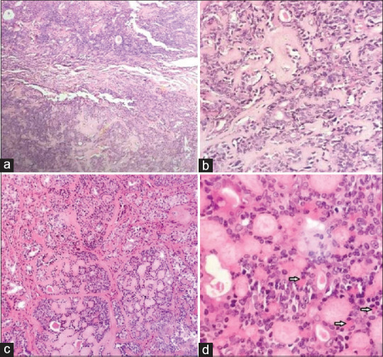
Pleomorphic adenoma is the most common benign salivary gland tumor deriving its name from varied morphological patterns in histopathology. The presence of chondromyxoid stroma in histopathology is characteristic of pleomorphic adenoma. Cellular variants without characteristic chondromyxoid stroma are rare and often pose a diagnostic challenge. We report a case of pleomorphic adenoma involving minor salivary glands of the palate presenting with a predominantly cellular histopathology. Immunohistochemical workup was pivotal in the diagnosis of this challenging case.
Keywords: Minor salivary glands; moderately cellular; palate; pleomorphic adenoma; salivary gland tumor.
Copyright: © 2021 Journal of Oral and Maxillofacial Pathology.
Conflict of interest statement
There are no conflicts of interest.
Figures


References
-
- Rito M, Fonesca I. Salivary gland neoplasms: Does morphologic diversity reflect tumor heterogeneity. Pathobiology. 2018;85:85–95. - PubMed
-
- Seethala RR. Salivary gland tumors: Current concepts and controversies. Surg Pathol Clin. 2017;10:155–76. - PubMed
-
- Ogle OE. Salivary gland diseases. Dent Clin North Am. 2020;64:87–104. - PubMed
-
- Pérez-de-Oliveira ME, Leonel AC, de Castro JF, Carvalho EJ, Vargas PA, Perez DE. Histopathological findings of intraoral pleomorphic adenomas: A retrospective study of a case series. Int J Surg Pathol. 2019;27:729–35. - PubMed
Publication types
LinkOut - more resources
Full Text Sources