

Expression of p53 at invasive front of oral squamous cell carcinoma and negative histopathological surgical margins to establish correlation at 3-year survival
- PMID: 33967511
- PMCID: PMC8083429
- DOI: 10.4103/jomfp.JOMFP_106_20
Expression of p53 at invasive front of oral squamous cell carcinoma and negative histopathological surgical margins to establish correlation at 3-year survival
Abstract
Background: Oral squamous cell carcinoma (OSCC) is the most common malignancy of the oral cavity. The histologic features of OSCC differ from area to area within same tumor, and most prognostic information can be revealed from the invasive front of tumor. The most accepted line of treatment is radical neck dissection. The boundaries of a resected specimen are the surgical margins (SMs), which are excised by the surgeon. The survival outcome is based on the status of these resected SMs. To avoid local recurrence and improve overall survival, it is necessary to attain negative SM. Apart from routine histopathology, the molecular assessment of resected margins has recently gained value which has a promising role for margin surveillance. The value of the use of molecular markers in the routine examination of resection specimens of OSCC has not yet established. It is crucial to identify the percentage of altered cells in SMs which go undetectable in the routine histopathology. It is essential to assess their role in recurrence and survival.
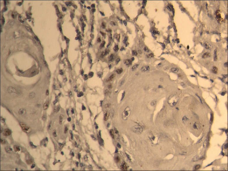
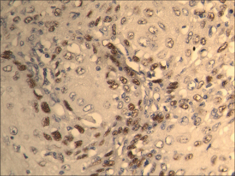
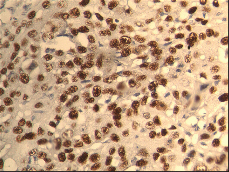
Materials and methods: The study was divided into two groups, i.e., Group I (control group): ten cases of normal oral mucosa and Group II consisted of thirty cases, in which biopsy samples of invasive tumor front and histopathologically negative SM of OSCC were included. Both the groups were subjected to p53 immunohistochemical staining.
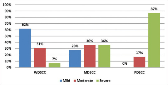
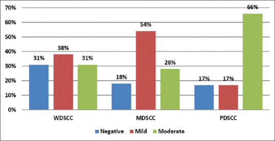
Results: There was overexpression of p53 at the deep tumor invasive front of OSCC associated with different histologic grades of malignancy.
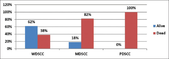
Conclusion: The overexpression of p53 at the invading tumor front with clear SMs is associated with poor survival. p53 expression at the tumor front can be a prognostic marker for OSCC.
Keywords: Oral squamous cell carcinoma; p53; survival; tumor-free margins.
Copyright: © 2021 Journal of Oral and Maxillofacial Pathology.
Conflict of interest statement
There are no conflicts of interest.
Figures
References
-
- Kumar V, Abbas AK, Fausto N. Neoplasia (Chapter 6) In: Kumar V, Abbas AK, Fausto N, editors. Robbins and Cotran Pathologic basis of Diseases. 7th ed. Philadelphia PA: Saunders; 2004. pp. 302–3.
-
- Cutilli T, Leocata P, Dolo V, Altobelli E. Evaluation of p53 protein as a prognostic factor for oral cancer surgery. Br J Oral Maxillofac Surg. 2013;51:922–7. - PubMed
LinkOut - more resources
Full Text Sources
Research Materials
Miscellaneous