

Osteosarcoma of jaws: Challenges in diagnosis
- PMID: 33967520
- PMCID: PMC8083400
- DOI: 10.4103/jomfp.JOMFP_142_20
Osteosarcoma of jaws: Challenges in diagnosis
Abstract
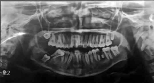
Osteosarcoma (OS) accounts for about 20% of all sarcomas with gnathic involvement seen in about 6%-10% of all OSs. The clinical presentation of OSs in the jaws is different from that of long bones as swelling is the most common complaint in patients with jaw OS followed by pain. The histopathologic variables seen are more favorable in OSs of jaws. Low-grade tumors are Stage I, high-grade tumors are Stage II and metastatic tumors (regardless of grade) are Stage III. A 17-year-old male patient reported with a complaint of the presence of an intra-oral growth gradually increasing in size in the right buccal mucosa region soft tissue enveloping the occlusal aspect of the right mandibular second molar. Extraorally swelling was present on the right side of the face for 4 months. Radiographically, there was a radiolucency from the distal aspect of right Mandibular second molar extending into the ramus region of the mandible with ill-defined borders. Hemi-mandibulectomy was done with the removal of the right mandible from the premolar region to condyle and coronoid processes. Microscopic evaluation of the sections after hematoxylin and eosin staining revealed interlacing fascicles of spindle-shaped cells arranged in a biphasic pattern and some areas of attempted bone formation evident in deeper sections. Tumor was an osteoblastic variety consisting of tumor osteoid surrounded by bizarrely arranged fibroblast-like cells. It showed positive staining with α-smooth muscle actin and Vimentin, suggesting a malignant tumor of mesenchymal cells with high myofibroblastic activity. Our case had small-cell histology; therefore, differential diagnosis was important.
Keywords: Chondroblastic; fibrosarcoma; osteoblastic; osteosarcoma; small-cell type osteosarcoma.
Copyright: © 2021 Journal of Oral and Maxillofacial Pathology.
Conflict of interest statement
There are no conflicts of interest.
Figures


References
-
- ElKordy MA, ElBaradie TS, ElSebai HI, Khairalla SM, Amin AA. Corrigendum to “Osteosarcoma of the jaw: Challenges in the diagnosis and treatment” [J Egypt Natl Cancer Inst (2018) 7–11] J Egypt Natl Canc Inst. 2018;30:123. - PubMed
-
- Lee RJ, Arshi A, Schwartz HC, Christensen RE. Characteristics and prognostic factors of osteosarcoma of the jaws. JAMA Otolaryngol Head Neck Surg. 2015;141:470. - PubMed
-
- Nagamine E, Hirayama K, Matsuda K, Okamoto M, Ohmachi T, Kadosawa T, et al. Diversity of histologic patterns and expression of cytoskeletal proteins in canine skeletal osteosarcoma. Vet Pathol. 2015;52:977–84. - PubMed
-
- Gupta S, Parikh S, Goel S. Parosteal osteosarcoma of mandible: A rare case report. J Cancer Res Ther. 2018;14:471–4. - PubMed
-
- Honoki K, Weiss K. Osteosarcoma: Biology, behavior and mechanisms. BoD – Books on Demand: Chapter-12. 2017. pp. 197–220. http://dx.doi.org/10.5772/67564 .
Publication types
LinkOut - more resources
Full Text Sources