

Sclerosing polycystic adenosis of minor salivary glands: Report of a rare case with diagnostic approach and review of literature
- PMID: 33967523
- PMCID: PMC8083427
- DOI: 10.4103/jomfp.JOMFP_186_20
Sclerosing polycystic adenosis of minor salivary glands: Report of a rare case with diagnostic approach and review of literature
Abstract
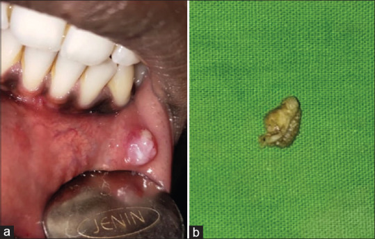
Sclerosing polycystic adenosis (SPA) is an uncommon entity occurring in the salivary glands, with majority of the cases reported in major salivary glands reminiscent of fibrocystic disease of the breast. SPA arising in minor salivary glands of the oral cavity constitutes an exceedingly rare phenomenon. Here, we report a case of SPA that presented as a solitary, submucosal mass on the left lower labial mucosa in a 19-year-old male. The pathology features and a clinicopathologic diagnostic approach highlighting key features are discussed here. Similar cases published in the English literature are reviewed.
Keywords: Mouth; salivary glands; sclerosing polycystic adenosis.
Copyright: © 2021 Journal of Oral and Maxillofacial Pathology.
Conflict of interest statement
There are no conflicts of interest.
Figures


Similar articles
-
Sclerosing Polycystic Adenosis of Hard Palate: A Rare Entity in Salivary Glands.Contemp Clin Dent. 2019 Oct-Dec;10(4):676-678. doi: 10.4103/ccd.ccd_94_19. Contemp Clin Dent. 2019. PMID: 32792830 Free PMC article.
-
Sclerosing polycystic adenosis of the buccal mucosa.Head Neck Pathol. 2008 Mar;2(1):31-5. doi: 10.1007/s12105-008-0042-9. Epub 2008 Feb 26. Head Neck Pathol. 2008. PMID: 20614339 Free PMC article. Review.
-
Sclerosing Polycystic Adenosis of Tongue.Bull Tokyo Dent Coll. 2018;59(2):121-125. doi: 10.2209/tdcpublication.2017-0029. Bull Tokyo Dent Coll. 2018. PMID: 29962419
-
Sclerosing polycystic adenosis of lower lip: A new and rare salivary gland entity.J Oral Maxillofac Pathol. 2018 May-Aug;22(2):263-265. doi: 10.4103/jomfp.JOMFP_254_17. J Oral Maxillofac Pathol. 2018. PMID: 30158783 Free PMC article.
-
Sclerosing polycystic adenosis of minor salivary glands: report of three cases and review of the literature.Oral Surg Oral Med Oral Pathol Oral Radiol Endod. 2007 Oct;104(4):516-20. doi: 10.1016/j.tripleo.2006.08.033. Oral Surg Oral Med Oral Pathol Oral Radiol Endod. 2007. PMID: 17150380 Review.
Cited by
-
Histopathological variations of Sclerosing Polycystic Adenoma: A systematic review.J Taibah Univ Med Sci. 2025 Jun 21;20(3):376-383. doi: 10.1016/j.jtumed.2025.06.005. eCollection 2025 Jun. J Taibah Univ Med Sci. 2025. PMID: 40612964 Free PMC article. Review.
-
Case of labial sclerosing polycystic adenoma with ductal carcinoma in situ (DCIS).BMJ Case Rep. 2021 Aug 17;14(8):e243736. doi: 10.1136/bcr-2021-243736. BMJ Case Rep. 2021. PMID: 34404657 Free PMC article.
References
-
- Westra WH, Kronz JD, Eisele DW. The impact of second opinion surgical pathology on the practice of head and neck surgery: A decade experience at a large referral hospital. Head Neck. 2002;24:684–93. - PubMed
-
- Swelam WM. The pathogenic role of Epstein-Barr virus (EBV) in sclerosing polycystic adenosis. Pathol Res Pract. 2010;206:565–71. - PubMed
-
- Gnepp DR, Wang LJ, Brandwein-Gensler M, Slootweg P, Gill M, Hille J. Sclerosing polycystic adenosis of the salivary gland: A report of 16 cases. Am J Surg Pathol. 2006;30:154–64. - PubMed
Publication types
LinkOut - more resources
Full Text Sources