

Synaptic changes and the response of microglia in a light-induced photoreceptor degeneration model
- PMID: 33967574
- PMCID: PMC8100860
Synaptic changes and the response of microglia in a light-induced photoreceptor degeneration model
Abstract
Purpose: To explore synaptic changes and the response of microglia in a light-induced photoreceptor degeneration model.
Methods: Sprague-Dawley rats were euthanized 1 h, 1 day, 3 days, 7 days, and 14 days after being exposed to intense blue light for 24 h. Hematoxylin and eosin (H&E) and terminal deoxynucleotidyl transferase dUTP nick-end labeling (TUNEL) staining were used to evaluate changes in the outer nuclear layer (ONL). Transmission electron microscopy (TEM) was applied to observe the ultrastructural changes in the synapses between the photoreceptors and second-order neurons. Western blotting was conducted to evaluate specific proteins, including postsynaptic density-95 (PSD-95), metabotropic glutamate receptor 6 (mGluR6), synapsin I, and synaptophysin. Immunofluorescence of CD11b and PKC-α or mGluR6 was used to explore the spatial relationships between microglial processes and synaptic elements. Immunoelectron microscopy of PSD-95 was performed to further confirm its engulfment of synaptic materials.
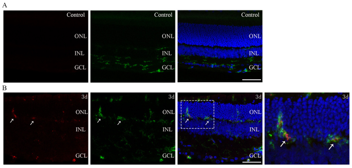
Results: H&E and TUNEL staining showed that the thickness of the ONL decreased markedly, and the number of apoptotic photoreceptors peaked at day 1. TEM revealed darkened photoreceptor terminals and that ribbons of them were floating in the cytoplasm, coinciding with the downregulation of PSD-95 and mGluR6. Downstream synaptic protein synapsin I and synaptophysin exhibited upregulation in the inner plexiform layer. Activated microglia migrated to the outer retina, and their processes were found in close proximity to synapses in the outer plexiform layer under light and electron microscopy levels. Double immunostaining of CD11b and mGluR6 showed colocalization. PSD-95-immunoreactive electron-dense materials were observed inside the microglia suggesting engulfment of synaptic components.
Conclusions: The study showed that there are early synaptic impairment and late compensatory changes in downstream synapses in this photic injury model. Activated microglia touched and directly engulfed synaptic materials. Microglia may play a role or a partial role in synaptic changes.
Copyright © 2021 Molecular Vision.
Figures


References
-
- Marigo V. Programmed cell death in retinal degeneration: targeting apoptosis in photoreceptors as potential therapy for retinal degeneration. Cell Cycle. 2007;6:652–5. - PubMed
-
- Santos A, Humayun MS, de Juan E, Jr, Greenburg RJ, Marsh MJ, Klock IB, Milam AH. Preservation of the inner retina in retinitis pigmentosa: a morphometric analysis. Arch Ophthalmol. 1997;115:511–5. - PubMed
-
- Nagar S, Krishnamoorthy V, Cherukuri P, Jain V, Dhingra NK. Early remodeling in an inducible animal model of retinal degeneration. Neuroscience. 2009;160:517–29. - PubMed
Publication types
MeSH terms
Substances
LinkOut - more resources
Full Text Sources
Research Materials
