. 2021 Mar 30;18(11):2285-2293.
doi: 10.7150/ijms.56198. eCollection 2021.

Anti-inflammatory effects of Flos Lonicerae Japonicae Water Extract are regulated by the STAT/NF-κB pathway and HO-1 expression in Virus-infected RAW264.7 cells

Affiliations

Anti-inflammatory effects of Flos Lonicerae Japonicae Water Extract are regulated by the STAT/NF-κB pathway and HO-1 expression in Virus-infected RAW264.7 cells

Hui-Wen Lin et al. Int J Med Sci. .

Abstract

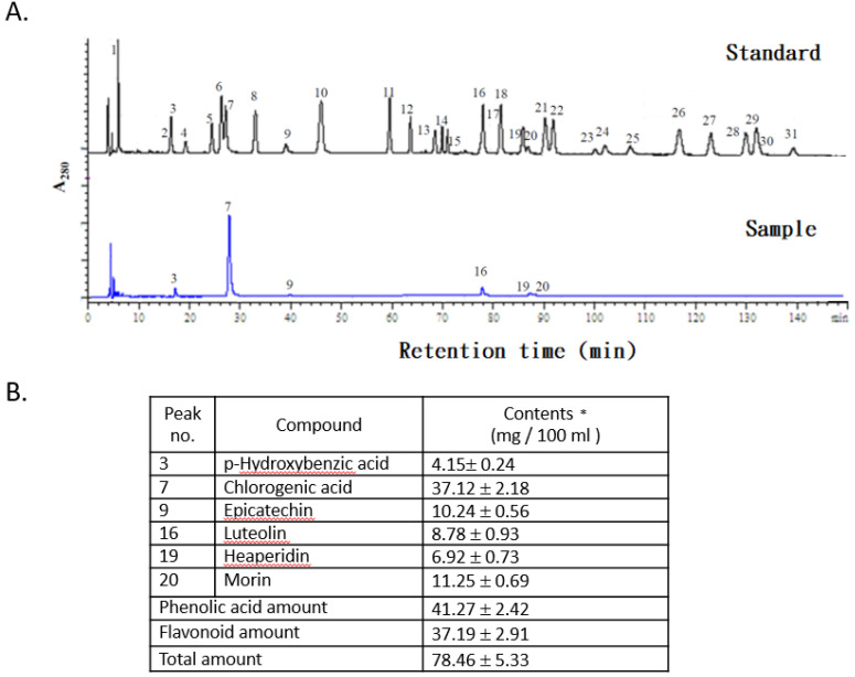
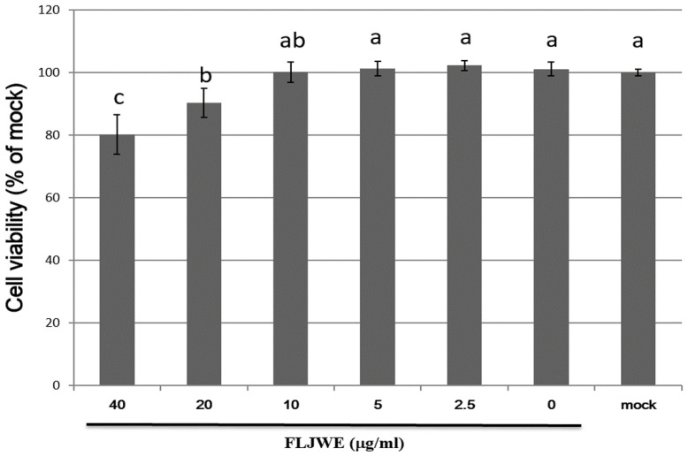
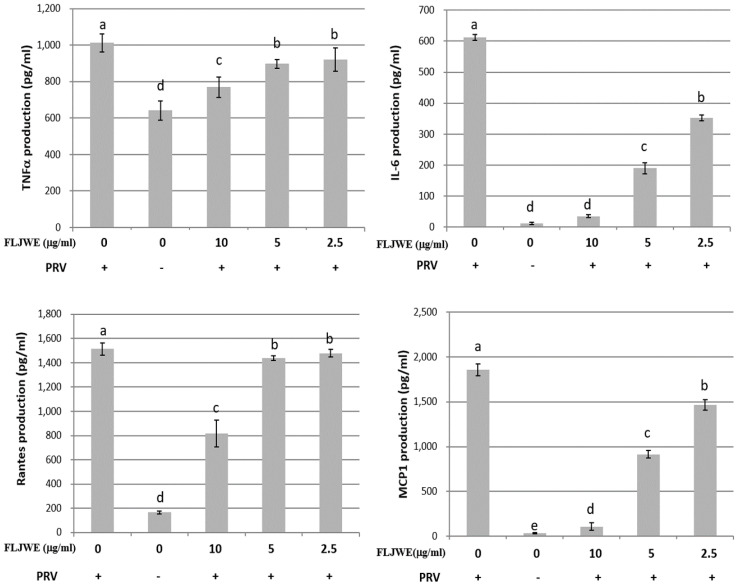
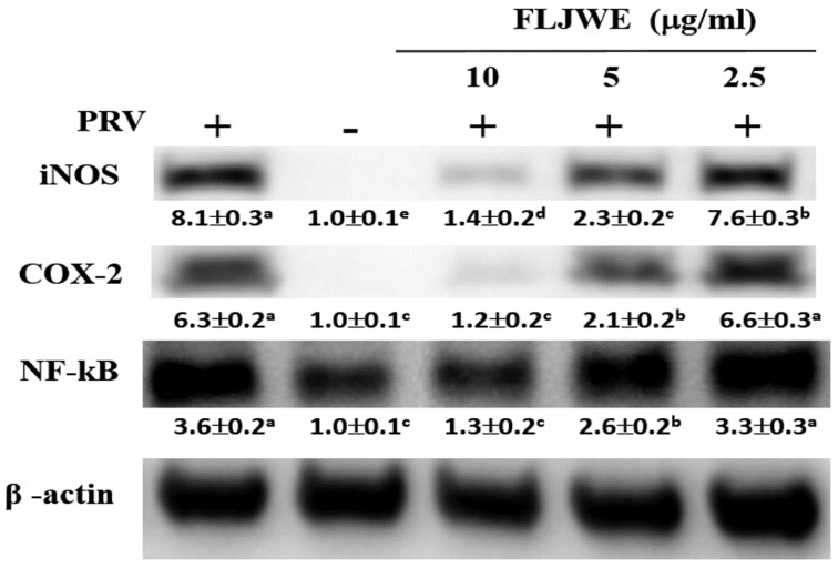
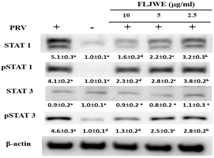
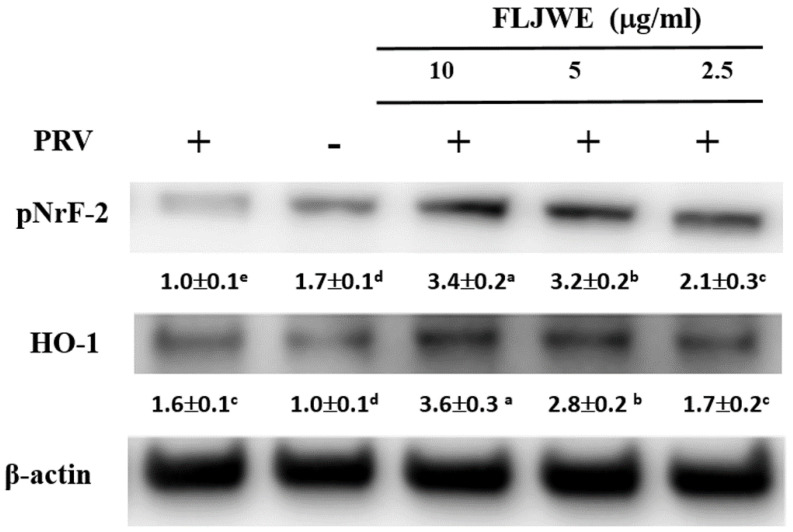
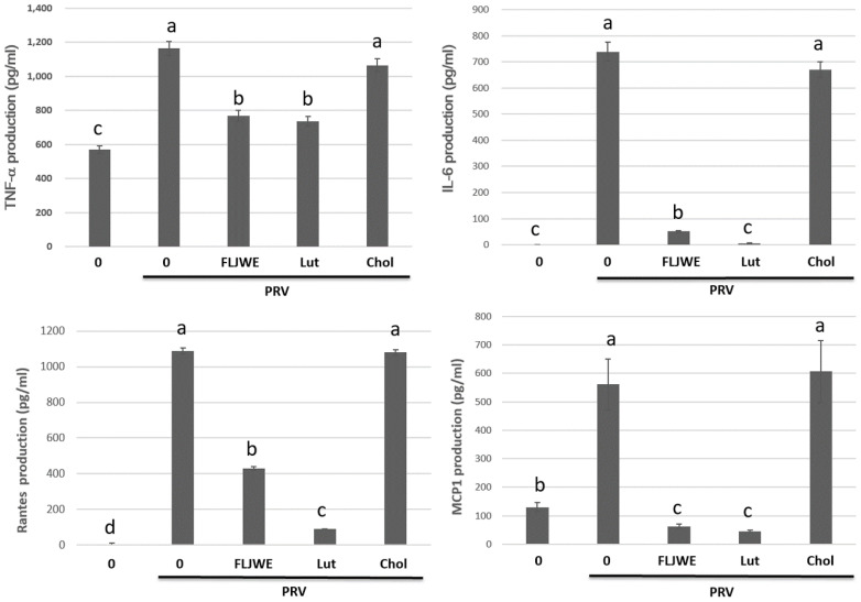
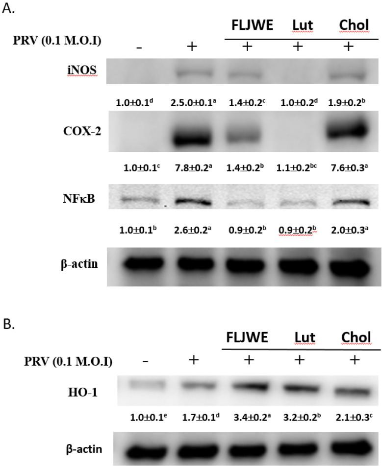
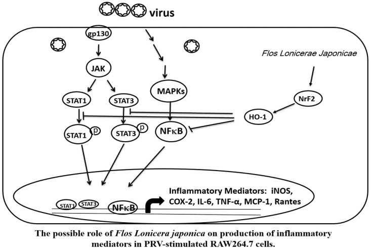
This study examined the effect of the Flos Lonicerae Japonicae water extract (FLJWE), chlorogenic acid, and luteolin on pseudorabies virus (PRV)-induced inflammation in RAW264.7 cells and elucidated related molecular mechanisms. The results revealed that FLJWE and luteolin, but not chlorogenic acid, inhibited the production of inducible nitric oxide synthase (iNOS), cyclooxygenase-2 (COX-2), and inflammatory cytokines in PRV-infected RAW 264.7 cells. We found that the FLJWE and luteolin suppressed nuclear factor (NF)-κB activation by inhibiting the phosphorylation of signal transducer and activator of transcription 1 and 3 (STAT1 and STAT3, respectively). Moreover, the FLJWE significantly upregulated the expression of pNrf2 and its downstream target gene heme oxygenase-1 (HO-1). Our data indicated that FLJWE and luteolin reduced the expression of proinflammatory mediators and inflammatory cytokines, such as COX-2 and iNOS, through the suppression of the JAK/STAT1/3-dependent NF-κB pathway and the induction of HO-1 expression in PRV-infected RAW264.7 cells. The findings indicate that the FLJWE can be used as a potential antiviral agent.

Keywords: Flos Lonicerae Japonicae water extract (FLJWE); antiviral inflammatory; heme oxygenase-1 (HO-1); pseudorabies virus (PRV).


Conflict of interest statement

Competing Interests: The authors have declared that no competing interest exists.

Figures

Figure 1
Figure 1
HPLC chromatograms of standards and the FLJWE. Peaks: (1) gallic acid, (2) gentisic acid, (3) p-hydroxybenzoic acid, (4) catechin, (5) vanillic acid, (6) caffeic acid, (7) chlorogenic acid, (8) syringic acid, (9) epicatechin, (10) p-coumaric acid, (11) ferulic acid, (12) sinapic acid, (13) p-anisic acid, (14) rutein, (15) quercitrin, (16) luteolin, (17) rosmaric acid, (18) neohesperidin, (19) hesperidin, (20) morin, (21) eriodictyol, (22) daidzein, (23) glycitrin, (24) quercetin, (25) diosmin, (26) naringenin (27) genistein, (28) nesperetin, (29) apigenin, (30) kaempferol, and (31) isorhamnetin. Data are presented as mean ± standard error of mean (n = 3).
Figure 2
Figure 2
Cell viability of RAW264.7 cells treated with various concentrations of the FLJWE. RAW264.7 cells were treated with various concentrations of FLJWE, and their cell viability was examined using an MTT assay. Data are presented as mean ± SD (n = 3). Mean values with different letters are significantly different (p < 0.05).
Figure 3
Figure 3
Inhibitory effects of the FLJWE on IL-6, MCP-1, RANTES, and TNF-α production in PRV-infected RAW264.7 cells. RAW264.7 cells were not treated or pretreated with various concentrations of the FLJWE (10, 5, and 2.5 μg/mL) for 1.5 h and then infected or not infected (control) with PRV (at an MOI of 0.1). After 24 h, the cultured medium was assayed to determine the levels of TNF-α, IL-6, RANTES, and MCP-1 through an enzyme-linked immunosorbent assay. The results are presented as mean ± SD (n = 3). Mean values with different letters are significantly different (p < 0.05).
Figure 4
Figure 4
Effect of the FLJWE on iNOS, COX-2, and NF-κB expression in PRV-infected RAW264.7 cells. RAW264.7 cells were not treated or pretreated with various concentrations of the FLJWE (2.5, 5, and 10 μg/mL) for 1.5 h and then infected or not infected (control) with PRV (at an MOI of 0.1) for 24 h. All proteins were subjected to 10% SDS-PAGE followed by Western blotting with iNOS, COX-2, NF-κB, and β-actin antibodies. Results are presented as mean ± SD (n = 3). Mean values with different letters are significantly different (p < 0.05).
Figure 5
Figure 5
Effect of FLJWE on the expression of the STAT1/3 pathway in PRV-infected RAW264.7 cells. RAW264.7 cells were not treated or pretreated with various concentrations of the FLJWE (2.5, 5, and 10 μg/mL) for 1.5 h and then infected (control) or not infected with PRV (at an MOI of 0.1) for 24 h. All proteins were subjected to 10% SDS-PAGE followed by Western blotting with STAT1/3, pSTAT1/3, and β-actin antibodies. Results are presented as mean ± SD (n = 3). Mean values with different letters are significantly different (p < 0.05).
Figure 6
Figure 6
Effect of FLJWE on the expression of HO-1 and pNrf2 in PRV-infected RAW264.7 cells. RAW264.7 cells were not treated or pretreated with various concentrations of the FLJWE (2.5, 5, and 10 μg/mL) for 1.5 h and then infected or not infected with PRV (at an MOI of 0.1) for 24 h. Total protein was subjected to 10% SDS-PAGE followed by Western blotting with HO-1, pNrf2, and β-actin antibodies. Results are presented as mean ± SD (n = 3). Mean values with different letters are significantly different (p < 0.05).
Figure 7
Figure 7
Effect of FLJWE, chlorogenic acid, and luteolin on inflammatory cytokines. RAW264.7 cells were not treated (control and PRV group) or pretreated with various concentrations of FLJWE, chlorogenic acid (Chol; 250 μM), or luteolin (Lut; 10 μM) for 1.5 h and then infected or uninfected (control) with PRV (at an MOI of 0.1). After 24 h, the cultured medium was assayed to determine the level of IL-6, MCP-1, RANTES, and TNF-α, which were measured using an enzyme-linked immunosorbent assay. Results are presented as mean ± SD (n = 3). Mean values with different letters are significantly different (p < 0.05).
Figure 8
Figure 8
Comparison of the effects of FLJWE, chlorogenic acid, and luteolin on iNOS, NF-κB, and HO-1 expression in PRV-infected RAW264.7 cells. RAW264.7 cells were not treated (control and PRV group) or pretreated with various concentrations of FLJWE, chlorogenic acid (Chol; 250 μM), or luteolin (Lut; 10 μM) for 1.5 h and then infected or not infected (control) with PRV for 24 h. All proteins were subjected to 10% SDS-PAGE followed by Western blotting with iNOS, NF-κB, HO-1, and β-actin antibodies. Mean values with different letters are significantly different (p < 0.05).
Figure 9
Figure 9
Possible role of Flos Lonicerae Japonicae in the production of inflammatory mediators in PRV-stimulated RAW264.7 cells.

References

    1. Li Y, Li W, Fu C. et al. Lonicerae japonicae flos and Lonicerae flos: a systematic review of ethnopharmacology, phytochemistry and pharmacology. Phytochem Rev. 2019;22:1–61. - PMC - PubMed
    1. Wang T, Yang B, Guan Q. et al. Transcriptional regulation of Lonicera japonica Thunb. during flower development as revealed by comprehensive analysis of transcription factors. BMC Plant Biology. 2019;19:198. - PMC - PubMed
    1. Cheng BC, Yu H, Su T. et al. A herbal formula comprising Rosae Multiflorae Fructus and Lonicerae Japonicae Flos inhibits the production of inflammatory mediators and the IRAK-1/TAK1 and TBK1/IRF3 pathways in RAW 264.7 and THP-1 cells. J Ethnopharmacol. 2015;174:195–9. - PubMed
    1. Kao ST, Liu CJ, Yeh CC. Protective and immunomodulatory effect of flos Lonicerae japonicae by augmenting IL-10 expression in a murine model of acute lung inflammation. J Ethnopharmacol. 2015;168:108–115. - PubMed
    1. Shang X, Pan H, Li M. et al. Lonicera japonica Thunb.: ethnopharmacology, phytochemistry and pharmacology of an important traditional Chinese medicine. J Ethnopharmacol. 2011;138:1–21. - PMC - PubMed

MeSH terms