. 2021 Apr 21:13:626974.
doi: 10.3389/fnagi.2021.626974. eCollection 2021.

Hippocampal and Hippocampal-Subfield Volumes From Early-Onset Major Depression and Bipolar Disorder to Cognitive Decline

Affiliations

Hippocampal and Hippocampal-Subfield Volumes From Early-Onset Major Depression and Bipolar Disorder to Cognitive Decline

Niels Hansen et al. Front Aging Neurosci. .

Abstract

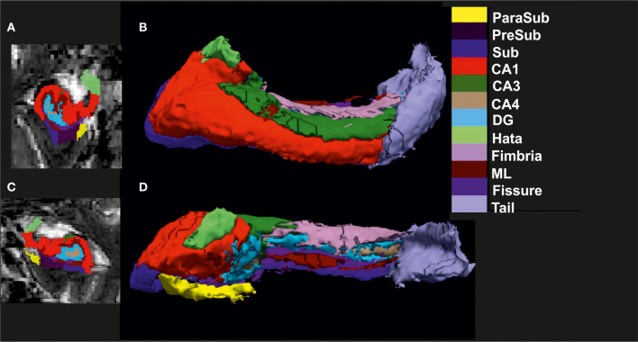
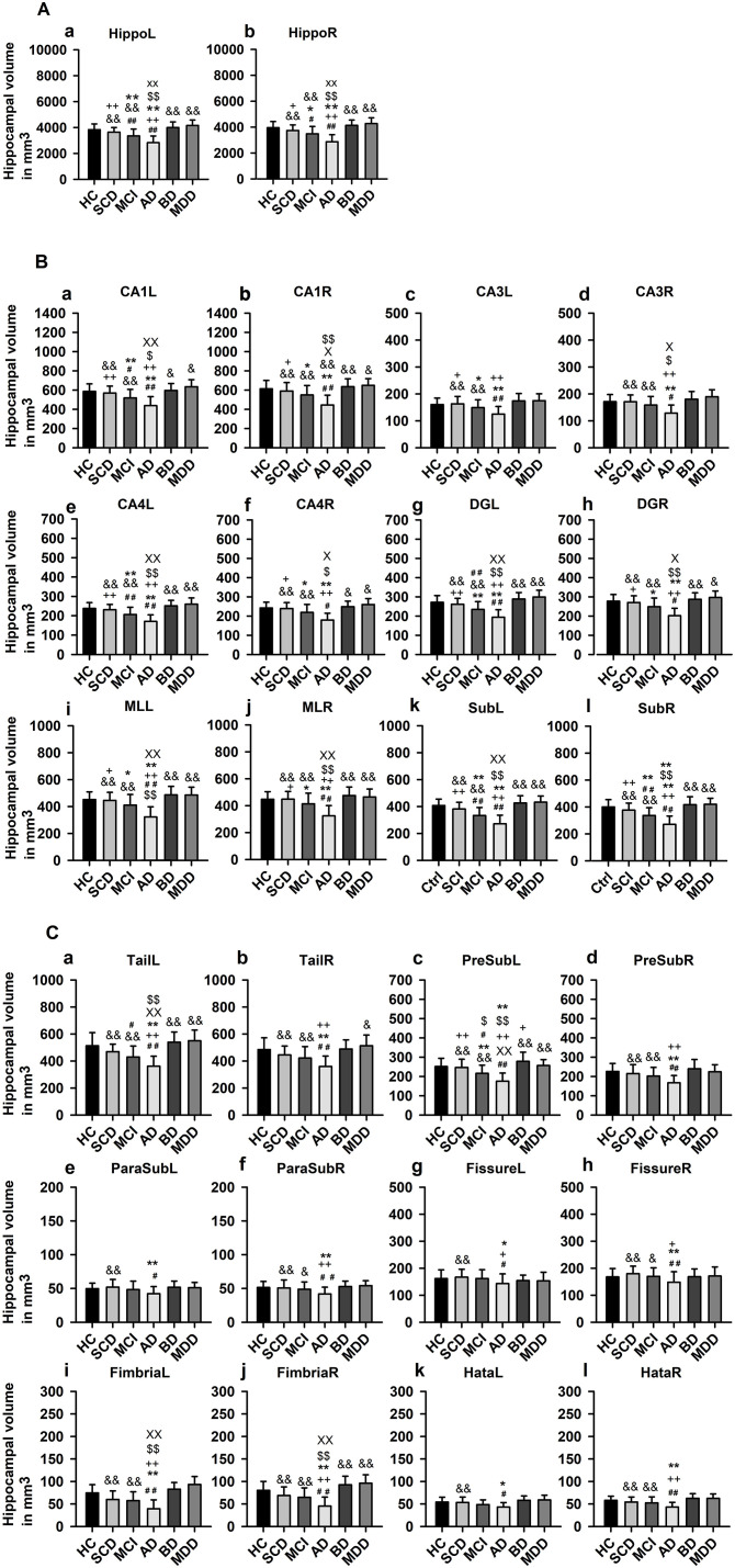
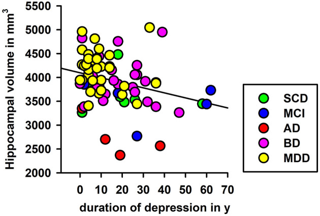
Background: The hippocampus and its subfields (HippSub) are reported to be diminished in patients with Alzheimer's disease (AD), bipolar disorder (BD), and major depressive disorder (MDD). We examined these groups vs healthy controls (HC) to reveal HippSub alterations between diseases. Methods: We segmented 3T-MRI T2-weighted hippocampal images of 67 HC, 58 BD, and MDD patients from the AFFDIS study and 137 patients from the DELCODE study assessing cognitive decline, including subjective cognitive decline (SCD), amnestic mild cognitive impairment (aMCI), and AD, via Free Surfer 6.0 to compare volumes across groups. Results: Groups differed significantly in several HippSub volumes, particularly between patients with AD and mood disorders. In comparison to HC, significant lower volumes appear in aMCI and AD groups in specific subfields. Smaller volumes in the left presubiculum are detected in aMCI and AD patients, differing from the BD group. A significant linear regression is seen between left hippocampus volume and duration since the first depressive episode. Conclusions: HippSub volume alterations were observed in AD, but not in early-onset MDD and BD, reinforcing the notion of different neural mechanisms in hippocampal degeneration. Moreover, duration since the first depressive episode was a relevant factor explaining the lower left hippocampal volumes present in groups.

Keywords: Alzheimer's disease; MRI volumetry; cognitive impairment; early-onset depression; hippocampal subfields; hippocampus.


Conflict of interest statement

The authors declare that the research was conducted in the absence of any commercial or financial relationships that could be construed as a potential conflict of interest.

Figures

Figure 1
Figure 1
Visualization of hippocampal subfield segmentation. (A) Left hippocampal subfields (HippSub) presented in a coronal MRI section, (B) Left HippSub illustrated in a 3D reconstruction, (C) Right HippSub presented in a coronal MRI section, and (D) Right HippSub illustrated in a 3D reconstruction. HippSub color code is on the right side of the figure. CA1/3/4, cornu ammonis 1/3/4; DG, granule cell layer-molecular layer of the dentate gyrus; Hata, hippocampus-amygdala transition area; ML, molecular layer; ParaSub, Parasubiculum; PreSub, Presubiculum; Sub, Subiculum.
Figure 2
Figure 2
Hippocampal subfield volumes across groups. (A) Whole hippocampus volumes compared in each hemisphere, (B) Hippocampal subfield (HippSub) volumes, including the CA1, CA3, CA4, DG, ML, and Sub as part of the hippocampus, compared in each hemisphere, and (C) Additional HippSub volumes including tail, PreSub, ParaSub, fissure, fimbria, Hata, compared in each hemisphere. Results refer to LSD post-hoc t-tests (two-sided) with Bonferroni correction between each condition. The significance level is indicated by different symbols: ##p < 0.005 vs. HC, **p < 0.005 vs. SCD, ++p < 0.005 vs. aMCI, &p < 0.005 vs. AD, $$p < 0.005 vs. BD, xxp < 0.005 vs. MDD, *p < 0.05 vs. SCD, +p < 0.05 vs. aMCI, &p < 0.05 vs. AD, $p < 0.05 vs. BD, xp < 0.05 vs. MDD. AD, Alzheimer's disease; BD, bipolar disorder; CA1/3/4, cornu ammonis 1/3/4; HC, healthy controls; DG, granule cell layer-molecular layer of the dentate gyrus; Hata, hippocampus-amygdala transition area; L, left; aMCI, amnestic mild cognitive impairment; MDD, major depressive disorder; ML, molecular layer; ParaSub, Parasubiculum; PreSub, Presubiculum; R, right; SCD, subjective cognitive decline; Sub, Subiculum.
Figure 3
Figure 3
Linear regression of depression duration and left hippocampal volumes. Significant regression analyses of depression duration and left hippocampal volume are shown. AD, Alzheimer's disease; BD, bipolar disorder; L, left; aMCI, amnestic mild cognitive impairment; MDD, major depressive disorder; SCD, subjective cognitive decline; y, years.

References

    1. Ang Y. S., Frontero N., Belleau E., Pizzagalli D. A. (2020). Disentangling vulnerability, state and trait features of neurocognitive impairments in depression. Brain 143, 3865–3877. 10.1093/brain/awaa314 - DOI - PMC - PubMed
    1. Ballmaier M., Narr K. L., Toga A. W., Elderkin-Thompson V., Thompson P. M., Hamilton L, et al. . (2008). Hippocampal morphology and distinguishing late-onset from early-onset elderly depression. Am. J. Psychiatry. 165, 229–237. 10.1176/appi.ajp.2007.07030506 - DOI - PMC - PubMed
    1. Berridge M. J. (2013). Dysregulation of neural calcium signaling in Alzheimer disease, bipolar disorder and schizophrenia. Prion 7, 2–13. 10.4161/pri.21767 - DOI - PMC - PubMed
    1. Brown E. M., Pierce M. E., Clark D. C., Fischl B. R., Iglesias J. E., Milberg W. P., et al. . (2020). Test-retest reliability of FreeSurfer automated hippocampal subfield segmentation within and across scanners Neuroimage 210:116563. 10.1016/j.neuroimage.2020.116563 - DOI - PubMed
    1. Brzezińska A., Bourke J., Rivera-Hernández R, Tsolaki M., Wozniak J., Kazmierski J. (2020). Depression in dementia or dementia in depression? systematic review of studies and hypotheses. Curr. Alzheimer Res. (2020) 17, 16–28. 10.2174/1567205017666200217104114 - DOI - PubMed

LinkOut - more resources