

Investigation of Extracellular Vesicles From SARS-CoV-2 Infected Specimens: A Safety Perspective
- PMID: 33968019
- PMCID: PMC8100059
- DOI: 10.3389/fimmu.2021.617042
Investigation of Extracellular Vesicles From SARS-CoV-2 Infected Specimens: A Safety Perspective
Abstract
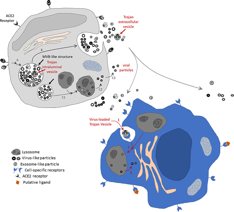
The coronavirus disease 2019 (COVID-19) pandemic, caused by the SARS-CoV-2 virus, is wreaking havoc around the world. Considering that extracellular vesicles (EVs) released from SARS-CoV-2 infected cells might play a role in a viremic phase contributing to disease progression and that standard methods for EV isolation have been reported to co-isolate viral particles, we would like to recommend the use of heightened laboratory safety measures during the isolation of EVs derived from SARS-CoV-2 infected tissue and blood from COVID-19 patients. Research needs to be conducted to better understand the role of EVs in SARS-CoV-2 infectivity, disease progression, and transmission. EV isolation procedures should include approaches for protection from SARS-CoV-2 contamination. We recommend the EV and virology scientific communities develop collaborative projects where relationships between endogenous EVs and potentially lethal enveloped viruses are addressed to better understand the risks and pathobiology involved.
Keywords: COVID-19; SARS-CoV-2; Trojan EV; Trojan exosome; extracellular vesicle; laboratory safety.
Copyright © 2021 Nunez Lopez, Casu and Pratley.
Conflict of interest statement
The authors declare that the research was conducted in the absence of any commercial or financial relationships that could be construed as a potential conflict of interest.
Figures
Similar articles
-
Extracellular Vesicles Analysis in the COVID-19 Era: Insights on Serum Inactivation Protocols towards Downstream Isolation and Analysis.Cells. 2021 Mar 4;10(3):544. doi: 10.3390/cells10030544. Cells. 2021. PMID: 33806297 Free PMC article.
-
COVID-19 and Extracellular Vesicles: An Intriguing Interplay.Kidney Blood Press Res. 2020;45(5):661-670. doi: 10.1159/000511402. Epub 2020 Sep 21. Kidney Blood Press Res. 2020. PMID: 32957112 Free PMC article. Review.
-
Proteomic profiling of single extracellular vesicles reveals colocalization of SARS-CoV-2 with a CD81/integrin-rich EV subpopulation in sputum from COVID-19 severe patients.Front Immunol. 2023 May 12;14:1052141. doi: 10.3389/fimmu.2023.1052141. eCollection 2023. Front Immunol. 2023. PMID: 37251406 Free PMC article.
-
Potential Therapeutic Effect of Micrornas in Extracellular Vesicles from Mesenchymal Stem Cells against SARS-CoV-2.Cells. 2021 Sep 12;10(9):2393. doi: 10.3390/cells10092393. Cells. 2021. PMID: 34572043 Free PMC article.
-
Potential therapeutic applications of extracellular vesicles in the immunopathogenesis of COVID-19.Pathol Res Pract. 2023 Jan;241:154280. doi: 10.1016/j.prp.2022.154280. Epub 2022 Dec 17. Pathol Res Pract. 2023. PMID: 36580795 Free PMC article. Review.
Cited by
-
Blood Nanoparticles - Influence on Extracellular Vesicle Isolation and Characterization.Front Pharmacol. 2021 Nov 10;12:773844. doi: 10.3389/fphar.2021.773844. eCollection 2021. Front Pharmacol. 2021. PMID: 34867406 Free PMC article. Review.
-
Mechanisms of COVID-19-induced kidney injury and current pharmacotherapies.Inflamm Res. 2022 Jan;71(1):39-56. doi: 10.1007/s00011-021-01520-8. Epub 2021 Nov 21. Inflamm Res. 2022. PMID: 34802072 Free PMC article. Review.
-
SARS-CoV-2-Derived RNA Fragment Induces Myocardial Dysfunction via siRNA-like Suppression of Mitochondrial ATP Synthase.Int J Mol Sci. 2025 Jun 4;26(11):5392. doi: 10.3390/ijms26115392. Int J Mol Sci. 2025. PMID: 40508201 Free PMC article.
-
Extracellular vesicle proteomics and phosphoproteomics identify pathways for increased risk in patients hospitalized with COVID-19 and type 2 diabetes mellitus.Diabetes Res Clin Pract. 2023 Mar;197:110565. doi: 10.1016/j.diabres.2023.110565. Epub 2023 Feb 1. Diabetes Res Clin Pract. 2023. PMID: 36736734 Free PMC article.
-
Comparison of viral inactivation methods on the characteristics of extracellular vesicles from SARS-CoV-2 infected human lung epithelial cells.J Extracell Vesicles. 2022 Dec;11(12):e12291. doi: 10.1002/jev2.12291. J Extracell Vesicles. 2022. PMID: 36468940 Free PMC article.
References
MeSH terms
Substances
LinkOut - more resources
Full Text Sources
Other Literature Sources
Medical
Miscellaneous