

Immune Checkpoints, a Novel Class of Therapeutic Targets for Autoimmune Diseases
- PMID: 33968036
- PMCID: PMC8097144
- DOI: 10.3389/fimmu.2021.645699
Immune Checkpoints, a Novel Class of Therapeutic Targets for Autoimmune Diseases
Abstract
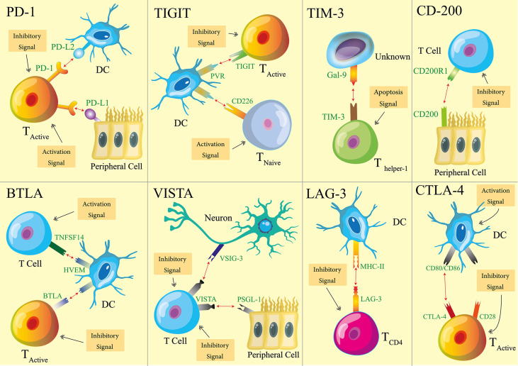
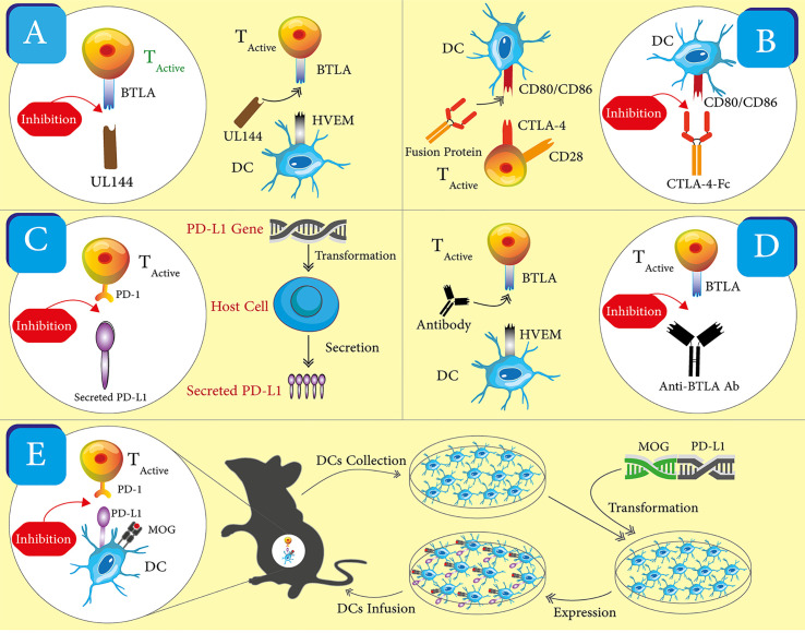
Autoimmune diseases, such as multiple sclerosis and type-1 diabetes, are the outcomes of a failure of immune tolerance. Immune tolerance is sustained through interplays between two inter-dependent clusters of immune activities: immune stimulation and immune regulation. The mechanisms of immune regulation are exploited as therapeutic targets for the treatment of autoimmune diseases. One of these mechanisms is immune checkpoints (ICPs). The roles of ICPs in maintaining immune tolerance and hence suppressing autoimmunity were revealed in animal models and validated by the clinical successes of ICP-targeted therapeutics for autoimmune diseases. Recently, these roles were highlighted by the clinical discovery that the blockade of ICPs causes autoimmune disorders. Given the crucial roles of ICPs in immune tolerance, it is plausible to leverage ICPs as a group of therapeutic targets to restore immune tolerance and treat autoimmune diseases. In this review, we first summarize working mechanisms of ICPs, particularly those that have been utilized for therapeutic development. Then, we recount the agents and approaches that were developed to target ICPs and treat autoimmune disorders. These agents take forms of fusion proteins, antibodies, nucleic acids, and cells. We also review and discuss safety information for these therapeutics. We wrap up this review by providing prospects for the development of ICP-targeting therapeutics. In summary, the ever-increasing studies and results of ICP-targeting of therapeutics underscore their tremendous potential to become a powerful class of medicine for autoimmune diseases.
Keywords: autoimmune diseases; cell; fusion protein; immune checkpoints; nucleic acid; therapeutics; viral protein.
Copyright © 2021 Zhai, Moosavi and Chen.
Conflict of interest statement
The authors declare that the research was conducted in the absence of any commercial or financial relationships that could be construed as a potential conflict of interest.
Figures
References
-
- Murphy K. Autoimmunity and transplantation. In: Janeway’s Immunobiology, 8th Edition. New York City, USA: Garland Science; (2012). p. 201.
-
- Lerner A, Jeremias P, Matthias T. The World Incidence and Prevalence of Autoimmune Diseases is Increasing. Int J Celiac Dis (2015) 3(4):151–5. 10.12691/ijcd-3-4-8 - DOI
Publication types
MeSH terms
Substances
Grants and funding
LinkOut - more resources
Full Text Sources
Other Literature Sources
Medical