Review
. 2021 Apr 22:12:639276.
doi: 10.3389/fpls.2021.639276. eCollection 2021.

Lipid Droplets in Unicellular Photosynthetic Stramenopiles

Affiliations
Review

Lipid Droplets in Unicellular Photosynthetic Stramenopiles

Nolwenn Guéguen et al. Front Plant Sci. .

Abstract

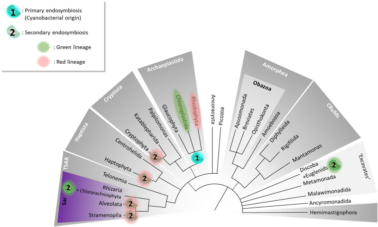
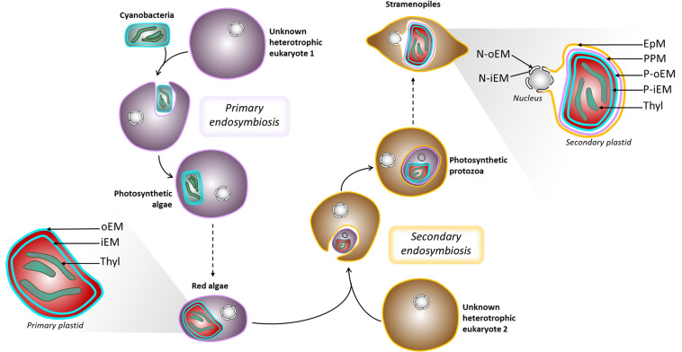
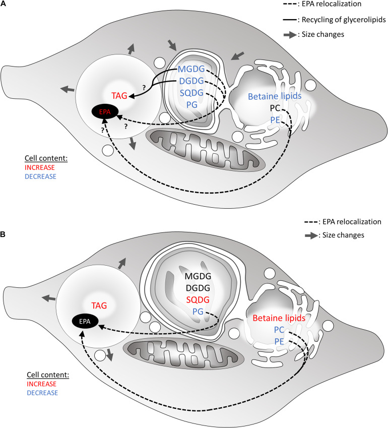
The Heterokonta or Stramenopile phylum comprises clades of unicellular photosynthetic species, which are promising for a broad range of biotechnological applications, based on their capacity to capture atmospheric CO2 via photosynthesis and produce biomolecules of interest. These molecules include triacylglycerol (TAG) loaded inside specific cytosolic bodies, called the lipid droplets (LDs). Understanding TAG production and LD biogenesis and function in photosynthetic stramenopiles is therefore essential, and is mostly based on the study of a few emerging models, such as the pennate diatom Phaeodactylum tricornutum and eustigmatophytes, such as Nannochloropsis and Microchloropsis species. The biogenesis of cytosolic LD usually occurs at the level of the endoplasmic reticulum. However, stramenopile cells contain a complex plastid deriving from a secondary endosymbiosis, limited by four membranes, the outermost one being connected to the endomembrane system. Recent cell imaging and proteomic studies suggest that at least some cytosolic LDs might be associated to the surface of the complex plastid, via still uncharacterized contact sites. The carbon length and number of double bonds of the acyl groups contained in the TAG molecules depend on their origin. De novo synthesis produces long-chain saturated or monounsaturated fatty acids (SFA, MUFA), whereas subsequent maturation processes lead to very long-chain polyunsaturated FA (VLC-PUFA). TAG composition in SFA, MUFA, and VLC-PUFA reflects therefore the metabolic context that gave rise to the formation of the LD, either via an early partitioning of carbon following FA de novo synthesis and/or a recycling of FA from membrane lipids, e.g., plastid galactolipids or endomembrane phosphor- or betaine lipids. In this review, we address the relationship between cytosolic LDs and the complex membrane compartmentalization within stramenopile cells, the metabolic routes leading to TAG accumulation, and the physiological conditions that trigger LD production, in response to various environmental factors.

Keywords: Microchloropsis; Nannochloropsis; Phaeodactylum; heterokont; lipid droplet (LD); seipin; stramenopile; triacylglycerol (TAG).


Conflict of interest statement

The authors declare that the research was conducted in the absence of any commercial or financial relationships that could be construed as a potential conflict of interest.

Figures

FIGURE 1
FIGURE 1
The Tree of Eukaryotes. The colored groupings correspond to currently defined “supergroups.” Unresolved branching orders among lineages are shown as multifurcations. Broken lines reflect minor uncertainties about the monophyly of certain groups. (1) Unique primary endosymbiosis event; (2) multiple secondary endosymbiosis events identified by Oborník (2019) as “complex endosymbioses.” The tree topology was adapted from Burki et al. (2020).
FIGURE 2
FIGURE 2
Schematic representation of plastid evolution. Schematic representation of primary and secondary endosymbiosis, and organelle architecture. EpM, epiplastidial membrane; PPM, periplastidial membrane; P-oEM and P-iEM, plastid outer and inner envelope membranes respectively; Thyl, thylakoids; N-oEM and P-iEM, nucleus outer and inner envelope membranes respectively.
FIGURE 3
FIGURE 3
Architecture of the membrane bound O-acyl transferase (MBOAT)-core of human diacylglycerol acyltransferase (DGAT1) enzyme (HsDGAT1). Adapted from Sui et al. (2020).
FIGURE 4
FIGURE 4
Different hypotheses regarding lipid droplet (LD) biogenesis in Phaeodactylum tricornutum. EpM, epiplastidial membrane; PpM, Periplastidial membrane; P-oEM, plastid outer envelope membrane; P-iEM, plastid inner envelope membrane; ER, endoplasmic reticulum; VN, vesicular network; SQDG, sulfoquinovosyldiacylglycerol; PC, phosphatidylcholine; DGTA, diacylglycerylhydroxymethyl-N,N,N-trimethyl-β-alanine; TAG, triacylglycerol). (A) Lipid droplets emerge from the EpM. The EpM membrane contains PC and specific DGTA species, but also SQDG. The abundance of C16:2 on the betaine lipid DGTA found in the lipid droplet monolayer (Lupette et al., 2019) supports a plastid emerging hypothesis as this fatty acid is major in the plastid but very minor in endomembranes (Abida et al., 2015). The origin of SQDG in the EpM is unknown and may be explained either by an export from the plastid or a synthetic pathway located in the EpM. (B) Lipid droplets form at an interface between the ER and the EpM. PC and DGTA can originate from both membranes, while SQDG comes from the EpM as described above. The peculiar DGTA composition (Lupette et al., 2019) suggests that only certain species are present at LD biogenesis sites although the sorting mechanism is unknown.
FIGURE 5
FIGURE 5
Hypothetical recycling of products of membrane glycerolipid breakdown for the formation of TAG in heterokonts subjected to a nitrogen or phosphate shortage. (A) Lipid remodeling under nitrogen starvation. Most glycerolipids are degraded, affecting the plastid size. (B) Lipid remodeling under phosphate starvation. In both scenarios, FA released from membrane lipids may be consumed via the β-oxidation pathway in the mitochondrion. Alternatively, down-products can be recycled to form TAG. Arrows show hypothetical conversions of membrane lipid down-products in the production of TAG, as well as in the increase in very long-chained PUFA (e.g., 20:5) in LDs. Glycerolipid conversions occur to rescue phospholipid degradation. DGDG, digalactosyldiacylglycerol; EPA, eicosapentaenoic acid; MGDG, monogalactosyldiacylglycerol; PC, phosphatidylcholine; PG, phosphatidylglycerol; SQDG, sulfoquinovosyldiacylglycerol; TAG, triacylglycerol.
FIGURE 6
FIGURE 6
Predicted structures of major lipid droplet proteins found in stramenopile models. Analysis of predicted structures and hydrophobic regions in (A–C) StLDP from Phaeodactylum tricornutum, (D–F) LDSP from Nannochloropsis s.l., and the highly similar (G–L) DOAP1 and PtLDP1 from Fistulifera solaris and P. tricornutum, respectively. (A,D,G) Secondary structure representations adapted from results obtained with Jpred4 prediction server (http://www.compbio.dundee.ac.uk/jpred/). Beta sheets are represented with red rectangles, and alpha helices with green arrows. (B,E,H) Predictions of transmembrane helices obtained with TMHMM prediction server (http://www.cbs.dtu.dk/services/TMHMM/). (C,F,I,J,K,L) Predicted tertiary structures obtained with Phyre2 (Protein Homology/analogy Recognition Engine V2.0) prediction server (http://www.sbg.bio.ic.ac.uk/∼phyre2/html/page.cgi?id = index) in ribbon diagrams (C,F,I,K,L), and surface representation (J). Obtained Phyre2 predictions span only a portion of the proteins, first and last amino acids (AAs) are specified in the figures. These regions are indicated in blue in the secondary structures representations (A,D,G) and TMHMM results (B,E,H). Hydrophobicity level of AAs is represented with a color code in (C,F,I,J), ranging from dark blue for very hydrophobic AAs, light blue for hydrophobic AAs, green for poorly hydrophobic or hydrophilic AAs, yellow for hydrophilic AAs, and red for very hydrophilic AAs. These predictions show that StLDP and LDSP have a hairpin structure, with the hairpin being enriched in hydrophobic residues. For DOAP1 and PtLDP1, a beta barrel structure bordered with two alpha helices is predicted. The membrane-spanning sequences of the beta barrel do not seem as enriched in hydrophobic residues as the alpha-helices of the hairpin structures of StLDP and LDSP, as already observed in the literature for these structures (Tamm et al., 2004; Wimley, 2009). The alpha helices potentially anchor the protein in the membrane as suggested by the TMHMM results. These alpha helices seen in the secondary structure are not present in the predicted tertiary structure due to incomplete models. To infer the position of the alpha helices regarding the beta barrel, first and last amino acids of the spanned regions are indicated with red arrows for one PtLDP1 model (K) and two possible DOAP1 models (L). DOAP1 model spanning the AAs 26–537 offers a more complete model than the model covering the AAs 26–462. However, a break in the predicted tertiary structure appears in this model between the two AAs indicated with blue arrows. The two alpha helices appear to be close to each other. However, it is not possible to conclude if the helices are in the same direction. AAs, amino acids; DOAP1, diatom oleosome-associated protein1; LDSP, lipid droplet surface protein; PtLDP1, P. tricornutum lipid droplet protein1; StLDP, stramenopile lipid droplet protein.

Similar articles

Cited by

References

    1. Abell B. M., Hahn M., Holbrook L. A., Moloney M. M. (2004). Membrane topology and sequence requirements for oil body targeting of oleosin: role of oleosin topology in oil body targeting. Plant J. 37 461–470. 10.1111/j.1365-313X.2003.01988.x - DOI - PubMed
    1. Abida H., Dolch L.-J., Meï C., Villanova V., Conte M., Block M. A., et al. (2015). Membrane glycerolipid remodeling triggered by nitrogen and phosphorus starvation in Phaeodactylum tricornutum. Plant Physiol. 167 118–136. 10.1104/pp.114.252395 - DOI - PMC - PubMed
    1. Adl S. N., Simpson A. G. B., Farmer M. A., Andersen R. A., Anderson R. O., Barta J. R., et al. (2005). The new higher level classification of eukaryotes with emphasis on the taxonomy of protists. J Eukaryot. Microbiol. 52 399–451. 10.1111/j.1550-7408.2005.00053.x - DOI - PubMed
    1. Alboresi A., Perin G., Vitulo N., Diretto G., Block M. A., Jouhet J., et al. (2016). Light remodels lipid biosynthesis in Nannochloropsis gaditana by modulating carbon partitioning between organelles. Plant Physiol. 171 2468–2482. 10.1104/pp.16.00599 - DOI - PMC - PubMed
    1. Alipanah L., Rohloff J., Winge P., Bones A. M., Brembu T. (2015). Whole-cell response to nitrogen deprivation in the diatom Phaeodactylum tricornutum. J. Exp. Bot. 66 6281–6296. 10.1093/jxb/erv340 - DOI - PMC - PubMed