. 2021 Jun;21(6):647.
doi: 10.3892/etm.2021.10079. Epub 2021 Apr 18.

Sinomenine activation of Nrf2 signaling prevents inflammation and cerebral injury in a mouse model of ischemic stroke

Affiliations

Sinomenine activation of Nrf2 signaling prevents inflammation and cerebral injury in a mouse model of ischemic stroke

Fangfang Bi et al. Exp Ther Med. 2021 Jun.

Abstract

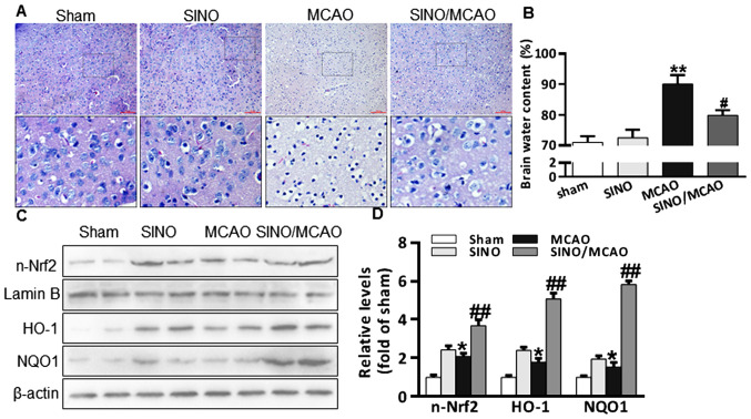
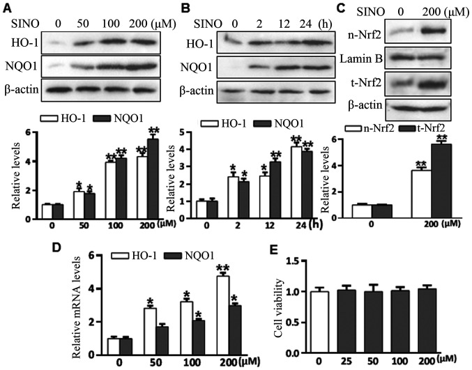
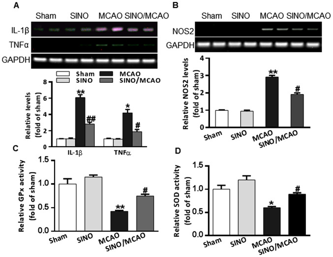
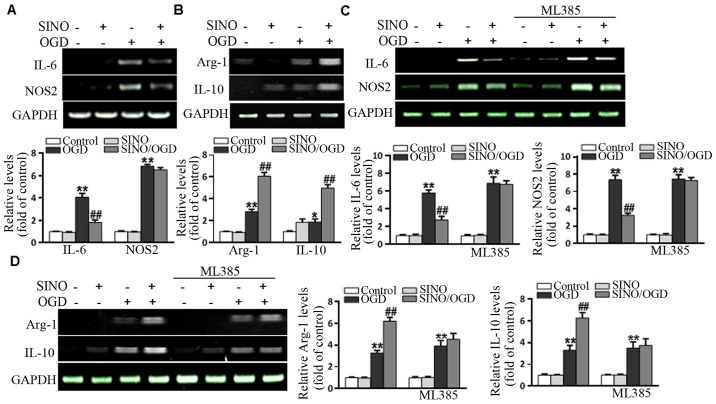
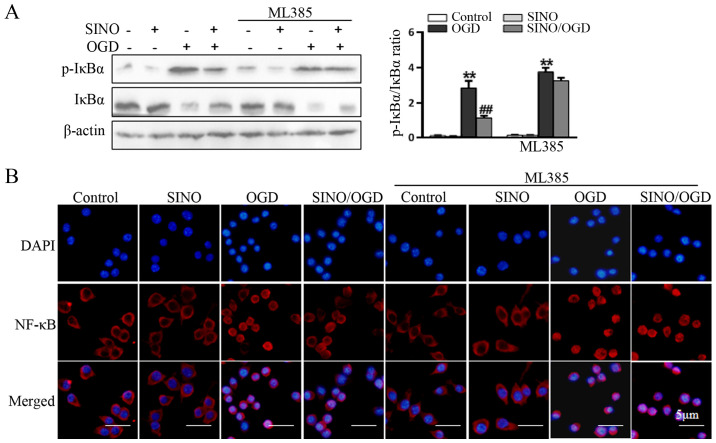
Sinomenine (SINO), which is used clinically to treat rheumatoid arthritis and neuralgia, is derived from the root and stems of Sinomenium acutum. SINO has been reported to exert analgesic, sedative and anti-inflammatory effects, and provides a protective role against shock and organ damage. Studies have suggested that SINO primarily exerts it anti-inflammatory function by inhibiting NF-κB signaling. There is also evidence to indicate that SINO may regulate inflammation Nuclear factor (erythroid-derived 2)-like 2 (Nrf2) signaling. The present study aimed to investigate whether the anti-inflammatory and cerebral protective effects of SINO were induced through Nrf2 both in vitro and in vivo. The results revealed that SINO significantly upregulated Nrf2 protein expression levels, increased Nrf2 nuclear translocation and the upregulated the protein expression levels of downstream factors. The treatment of a middle cerebral artery occlusion model mice with SINO effectively reduced cerebral damage and inflammation, and restored the balance in cerebral oxidative stress. In addition, SINO treatment also promoted Nrf2-dependent microglia M1/M2 polarization and inhibited the phosphorylation of IκBα as well as NF-κB nuclear translocation. This revealed an important upstream event that contributed to its anti-inflammatory and cerebral tissue protective effects. In conclusion, the findings of the present study identified a novel pathway through which SINO may exert its anti-inflammatory and cerebral protective functions, and provided a molecular basis for the potential applications of SINO in the treatment of cerebral inflammatory disorders.

Keywords: cerebral protection; inflammation; microglia; nuclear factor-erythroid 2-related factor; sinomenine.


Conflict of interest statement

The authors declare that they have no competing interests.

Figures

Figure 1
Figure 1
SINO treatment relieves MCAO-induced cerebral injuries. (A) Representative micrographs of H&E staining of brain sections (x20 magnification; scale bar, 50 µm). (B) Brain water content analysis in brain tissues from Sham (sham-operated, n=6), SINO (treatment only, n=6), MCAO (MCAO-operated, n=6) and SINO/MCAO (MCAO operated plus SINO treatment, n=6). (C) Western blot analysis of n-Nrf2, HO-1 and NQO1 protein expression levels from brain tissue. The samples from two randomly selected brains in each group were presented. (D) Quantification of brain protein expression levels. All the experiments were repeated at least three times. The data are presented as the mean ± SEM. *P<0.05 and **P<0.01 vs. Sham; #P<0.05 and ##P<0.01 vs. MCAO. HO-1, heme oxygenase-1; MCAO, middle cerebral artery occlusion; n-Nrf2, nuclear-nuclear factor-erythroid 2-related factor; NQO1, NAD(P)H: Quinoneoxidoreductase 1; SINO, sinomenine.
Figure 2
Figure 2
SINO treatment activates the Nrf2 signaling pathway. (A) BV2 cells were treated with a serial of doses of SINO for 24 h. HO-1 and NQO1 protein expression levels were assayed using western blotting. (B) BV2 cells were treated with SINO (200 µM) for various durations. HO-1 and NQO1 protein expression levels were assayed using western blotting. (C) BV2 cells were treated with SINO (200 µM) for 4 h. n-Nrf2 and t-Nrf2 protein expression levels were assayed using western blotting. Nuclear protein Lamin B and β-actin were used as controls. (D) BV2 cells were treated with serial doses of SINO for 24 h. The mRNA expression levels of HO-1 and NQO1 were measured using reverse transcription-semi-quantitative PCR. (E) Cell viability assay. BV2 cells were treated with various doses of SINO for 24 h. The cell viability was assayed by a CCK-8 kit. All the experiments were repeated at least three times. *P<0.05 and **P<0.01 vs. Control HO-1, heme oxygenase-1; NQO1, NAD(P)H: Quinoneoxidoreductase 1; n-, nuclear; Nrf2; nuclear factor-erythroid 2-related factor; SINO, sinomenine; t-, total.
Figure 3
Figure 3
SINO treatment mitigates MCAO-associated inflammation and oxidative stress. (A) RT-sqPCR analysis of TNF-α and IL-1β mRNA expression levels in brain tissues. The results of two samples from each group were presented. (B) RT-sqPCR analysis of NOS2 mRNA expression levels in brain tissues. (C) The relative enzyme activities of GPx from the brain tissue of the four groups. (D) The relative enzyme activities of SOD from the brain tissue of four groups. All experiments were repeated at least three times. The data are presented as the mean ± SEM. *P<0.05 and **P<0.01 vs. Sham; #P<0.05 and ##P<0.01 vs. MCAO. GPx, glutathione peroxidase; MCAO, middle cerebral artery occlusion; NOS2, nitric oxide synthase 2; RT-sqPCR, reverse transcription-semi-quantitative PCR; SINO, sinomenine; SOD, superoxide dismutase.
Figure 4
Figure 4
SINO treatment regulates microglia polarization and inflammation in an Nrf2-dependent manner. BV2 cells were stimulated with OGD for 4 h followed by treatment with SINO (200 µM) for 12 h. The mRNA expression levels of (A) M1 markers (IL-6/NOS2) and (B) M2 markers (IL-10/Arg-1) were determined using RT-sqPCR. BV2 cells were pretreated with ML385 (5 µM) for 48 h to inhibit Nrf2 expression. Cells were then stimulated with OGD for 4 h followed by treatment with SINO (200 µM) for 12 h. The mRNA expression levels of (C) M1 and (D) M2 markers were measured using RT-sqPCR. The data are presented as the mean ± SEM of three independent experiments. *P<0.05 and **P<0.01 vs. Control; ##P<0.01 vs. OGD. Arg-1, arginase-1; NOS2, nitric oxide synthase 2; OGD, oxygen and glucose deprivation; SINO, sinomenine; RT-sqPCR, reverse transcription-semi-quantitative PCR.
Figure 5
Figure 5
SINO treatment regulates microglia inflammation in an Nrf2-dependent manner. (A) BV2 cells were pretreated with ML385 (5 µM) for 48 h to inhibit Nrf2 expression. Cells were then stimulated with OGD for 4 h followed by treatment with SINO (200 µM) for 12 h. The protein expression levels of p-IκBα and total IκBα were analyzed using western blotting. (B) Immuno-fluorescent staining of cells was performed using an NF-κB p65 primary antibody and Dylight488 conjugated secondary antibody (middle row of panels). The cells were also stained with DAPI (top row of panels) and merged with NF-κB images (lower row of panels). The data are presented as the mean ± SEM of three independent experiments. **P<0.01 vs. Control; ##P<0.01 vs. OGD. OGD, oxygen and glucose deprivation; p-, phosphorylated; SINO, sinomenine.

Similar articles

Cited by

References

    1. Jeon JH, Jung HW, Jang HM, Moon JH, Park KT, Lee HC, Lim HY, Sur JH, Kang BT, Ha J, Jung DI. Canine model of ischemic stroke with permanent middle cerebral artery occlusion: Clinical features, magnetic resonance imaging, histopathology, and immunohistochemistry. J Vet Sci. 2015;16:75–85. doi: 10.4142/jvs.2015.16.1.75. - DOI - PMC - PubMed
    1. Ito M, Shichita T, Okada M, Komine R, Noguchi Y, Yoshimura A, Morita R. Bruton's tyrosine kinase is essential for NLRP3 inflammasome activation and contributes to ischaemic brain injury. Nat Commun. 2015;6(7360) doi: 10.1038/ncomms8360. - DOI - PMC - PubMed
    1. Fu Y, Liu Q, Anrather J, Shi FD. Immune interventions in stroke. Nat Rev Neurol. 2015;11:524–535. doi: 10.1038/nrneurol.2015.144. - DOI - PMC - PubMed
    1. Pont-Lezica L, Bechade C, Belarif-Cantaut Y, Pascual O, Bessis A. Physiological roles of microglia during development. J Neurochem. 2011;119:901–908. doi: 10.1111/j.1471-4159.2011.07504.x. - DOI - PubMed
    1. Subhramanyam CS, Wang C, Hu Q, Dheen ST. Microglia-mediated neuroinflammation in neurodegenerative diseases. Semin Cell Dev Biol. 2019;94:112–120. doi: 10.1016/j.semcdb.2019.05.004. - DOI - PubMed