

Imaging Studies in Otosclerosis: An Up-to-date Comprehensive Review
- PMID: 33968239
- PMCID: PMC8096512
- DOI: 10.1055/s-0040-1715149
Imaging Studies in Otosclerosis: An Up-to-date Comprehensive Review
Abstract
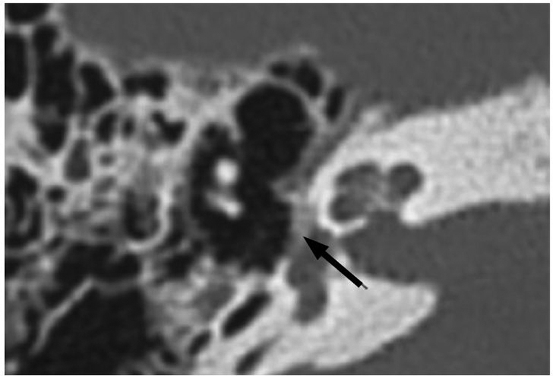
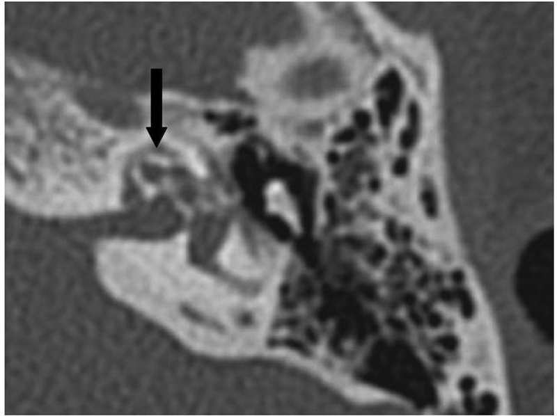
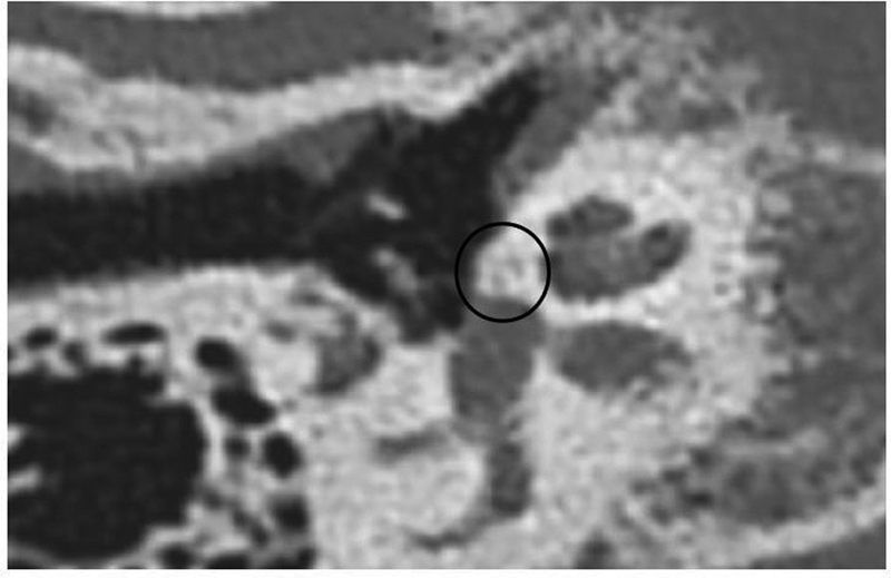
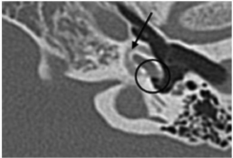
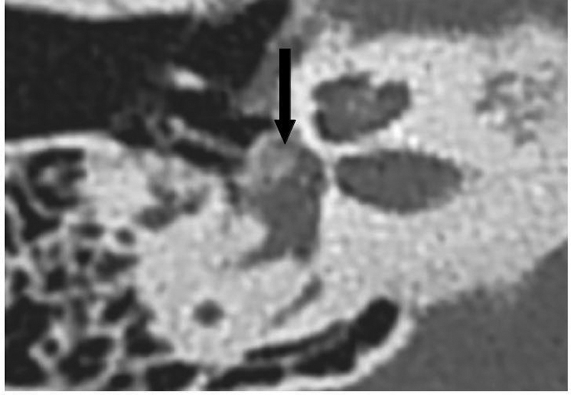
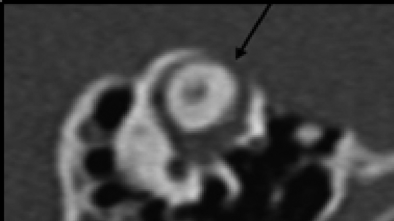
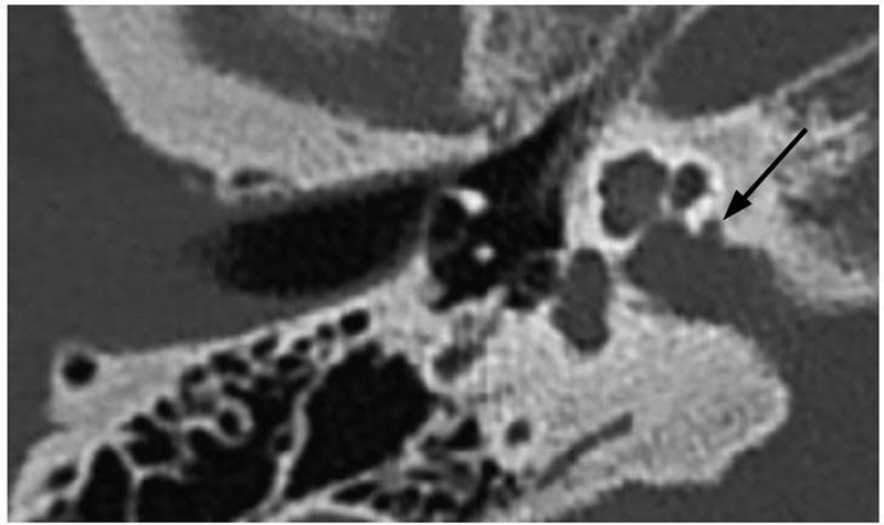
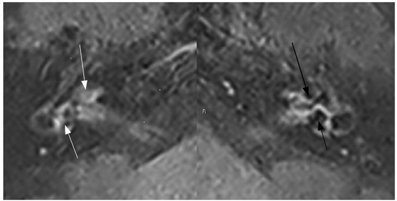
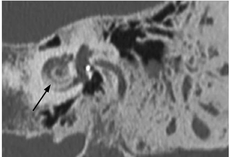
Introduction Otosclerosis is a primary osteodystrophy of the otic capsule, frequently responsible for acquired hearing loss in adults. Although the diagnostic value of imaging investigations in otosclerosis is debatable, they might still be employed with different goals within the context of the disease. Objectives The present paper aims to review the most recent literature on the use of imaging studies in otosclerosis for the most varied purposes, from routine application and differential diagnosis to prognostic prediction and investigation of surgical failure. Data Synthesis The diagnosis of otosclerosis is usually clinical, but computed tomography (CT) is paramount in particular cases for the differential diagnosis. The routine use, however, is not supported by strong evidence. Even so, there is growing evidence of the role of this method in surgical planning and prediction of postoperative prognosis. In specific scenarios, for example when superior semicircular canal dehiscence (SSCD) syndrome is suspected or in surgical failure, CT is crucial indeed. Magnetic resonance imaging (MRI), however, has limited - although important - indications in the management of individuals with otosclerosis, especially in the evaluation of postoperative complications and in the follow-up of medical treatment in active ostosclerosis. Conclusion Imaging studies have a broad range of well-established indications in otosclerosis. Besides, although the routine use of CT remains controversial, the most recent papers have shed light into new potential benefits of imaging prior to surgery.
Keywords: X-Ray; computed; diagnostic imaging; magnetic resonance imaging; otosclerosis; stapes surgery; tomography.
Fundação Otorrinolaringologia. This is an open access article published by Thieme under the terms of the Creative Commons Attribution-NonDerivative-NonCommercial License, permitting copying and reproduction so long as the original work is given appropriate credit. Contents may not be used for commecial purposes, or adapted, remixed, transformed or built upon. ( https://creativecommons.org/licenses/by-nc-nd/4.0/ ).
Figures
References
-
- Goh J PN, Chan L L, Tan T Y. MRI of cochlear otosclerosis. Br J Radiol. 2002;75(894):502–505. - PubMed
-
- Glasscock M, Shambaugh G. Philadelphia: WB Saunders; 1990. Surgery of the Ear . 4th ed .
LinkOut - more resources
Full Text Sources