. 2021 Apr 21:2021:5558687.
doi: 10.1155/2021/5558687. eCollection 2021.

Ulinastatin Ameliorates IL-1 β-Induced Cell Dysfunction in Human Nucleus Pulposus Cells via Nrf2/NF- κ B Pathway

Affiliations

Ulinastatin Ameliorates IL-1 β-Induced Cell Dysfunction in Human Nucleus Pulposus Cells via Nrf2/NF- κ B Pathway

Xi Luo et al. Oxid Med Cell Longev. .

Abstract

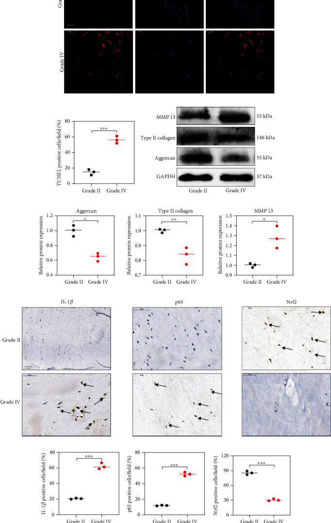
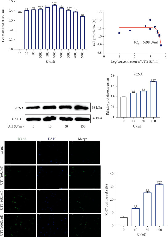
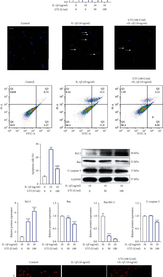
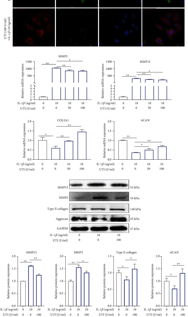
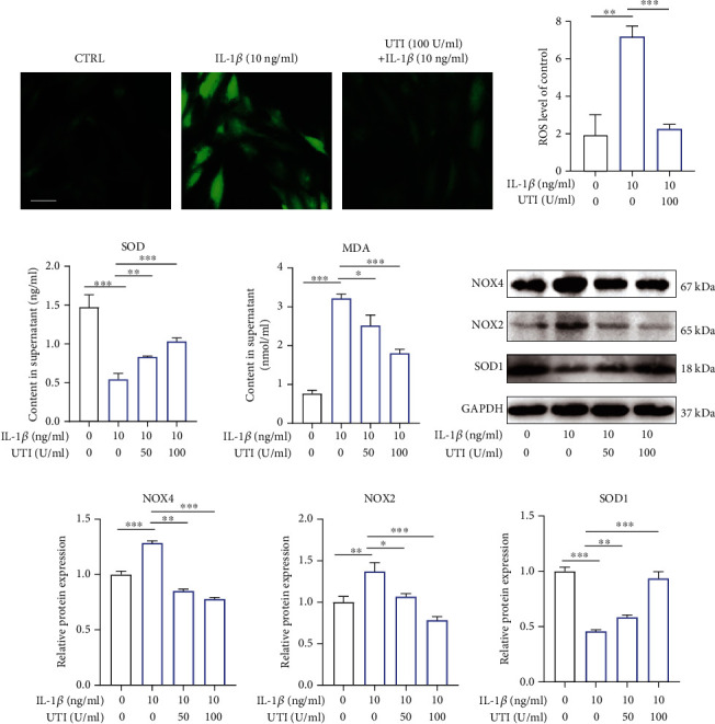
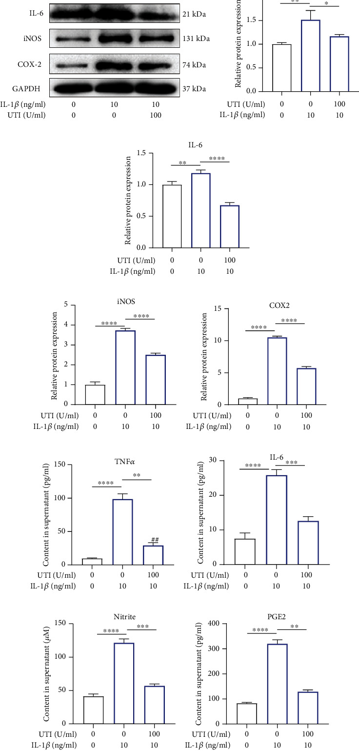
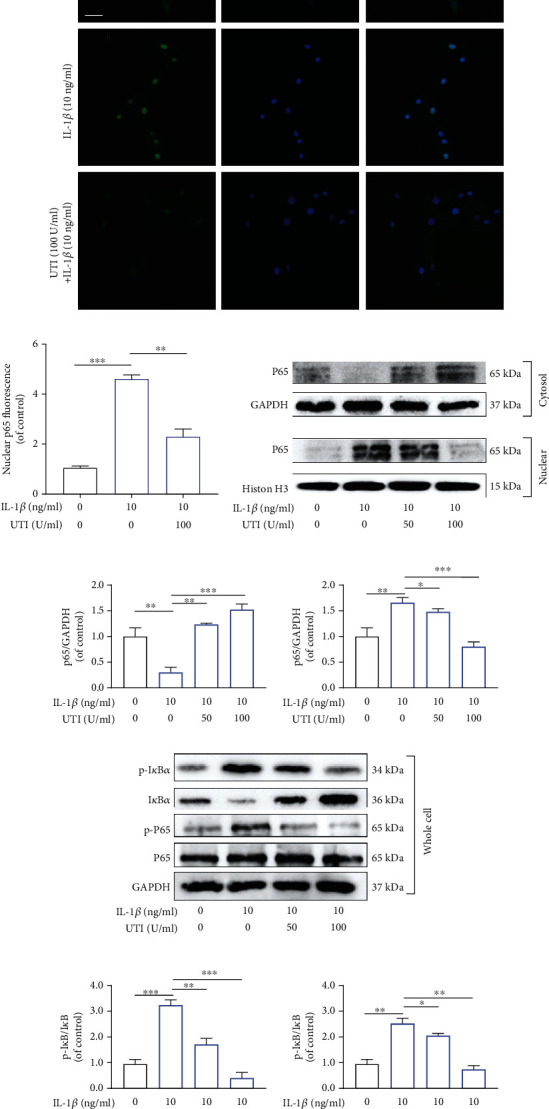
Low back pain (LBP) has been a wide public health concern worldwide. Among the pathogenic factors, intervertebral disc degeneration (IDD) has been one of the primary contributors to LBP. IDD correlates closely with inflammatory response and oxidative stress, involving a variety of inflammation-related cytokines, such as interleukin 1 beta (IL-1β), which could result in local inflammatory environment. Ulinastatin (UTI) is a kind of acidic protein extracted from human urine, which inhibits the release of tumor necrosis factor alpha (TNF-α) and other inflammatory factors to protect organs from inflammatory damage. However, whether this protective effect of UTI on human nucleus pulposus (NP) exists, and how UTI affects the biological behaviors of human NP cells during IDD remain elusive. In this current study, we revealed that UTI could improve the viability of NP cells and promote the proliferation of NP cells. Additionally, UTI could protect human NP cells via ameliorating IL-1β-induced apoptosis, inflammatory response, oxidative stress, and extracellular matrix (ECM) degradation. Molecular mechanism analysis suggested that the protective effect from UTI on IL-1β-treated NP cells were through activating nuclear factor- (erythroid-derived 2-) like 2 (Nrf2)/heme oxygenase-1 (HO-1) signaling pathway and the suppression of NF-κB signaling pathway. Therefore, UTI may be a promising therapeutic medicine to ameliorate IDD.


Conflict of interest statement

The authors have declared no conflict of interest.

Figures

Figure 1
Figure 1
The protein expression of Nrf2 and p65 in human NP tissues. (a) The apoptotic NP cells (red) in intervertebral disc between Grade II and Grade IV were visualized via TUNEL staining and the nuclei were stained with DAPI. Scar bar = 300 μm. (b) Three randomized versions were selected, and TUNEL staining-positive cells were quantified via the amount of red fluorescence. (c, d) Protein bands and quantification of expression levels of Aggrecan, Type II collagen, and MMP 13. Scar bar = 100 μm. (e) Immunohistochemical results were used to examine the protein levels of IL-1β, p65, and Nrf2 in the human NP tissue with Grade II and Grade IV. (f) Three versions were randomly selected, and the stained cells were quantified separately. p < 0.05, ∗∗p < 0.01, ∗∗∗p < 0.001, ∗∗∗∗p < 0.0001.
Figure 2
Figure 2
Effects of UTI on the proliferative ability in human NP cells. (a) The biological effect of UTI on the cell viability of NP cells (n = 5). (b) IC50 of UTI for human NP cells. (c, d) Protein bands and quantification of expression levels of PCNA (n = 3). (e, f) Expression of Ki-67 (green) was detected by the immunofluorescence. The ratios of cells with green fluorescence were calculated. Scar bar = 200 μm. p < 0.05, ∗∗p < 0.01, ∗∗∗p < 0.001, ∗∗∗∗p < 0.0001.
Figure 3
Figure 3
Biological effect of UTI on IL-1β-induced apoptosis in human NP cells. (a, b) Protein bands and quantification of expression levels of Bcl-2, Bax, and cleaved caspase 3 in IL-1β-induced NP cells. Bax/Bcl-2 ratios were calculated (n = 3). (c) Effect of UTI with indicated concentrations on cell viability of IL-1β-induced NP cells (n = 5). (d) Hoechst staining was used to conduct analysis of the nuclear morphology. Scar bar = 100 μm. (e, f) The rates of apoptosis of human NP cells as determined by flow cytometry (n = 3). (g, h) Protein bands and quantification of expression levels of Bcl-2, Bax, and cleaved caspase 3 in IL-1β-induced NP cells cotreated with UTI. Bax/Bcl-2 ratios were calculated (n = 3). (i, j) The JC-1 monomers (red) and JC-1 aggregates (green) were detected by the fluorescent probe JC-1, and the ratios of JC-1 aggregate/monomer were calculated. Scar bar = 200 μm. p < 0.05, ∗∗p < 0.01, ∗∗∗p < 0.001, ∗∗∗∗p < 0.0001. indicated the comparison between group IL-1β and group control. #p < 0.05, ##p < 0.01, ###p < 0.001, ####p < 0.0001. #indicated the comparison between group IL-1β and group UTI+IL-1β.
Figure 4
Figure 4
UTI ameliorated IL-1β-induced ECM degradation in human NP cells. (a) The expression of MMP3 and type II collagen by the immunofluorescence. Scar bar = 50 μm. (b) qRT-qPCR was used to evaluate the mRNA expression of MMP 3, MMP 13, ACAN, and COL2A1 (n = 3). (c, d) Protein bands and quantification of protein levels of MMP 13, MMP 3, type II collagen, and aggrecan. GAPDH as an internal control (n = 3). (e) IL-1β-induced differential expression of MMP 13, MMP 3, type II collagen, and aggrecan were measured by ELISA cotreated with UTI in a dose-dependent manner in the cultural supernatant of NP cells (n = 3). p < 0.05, ∗∗p < 0.01, ∗∗∗p < 0.001, ∗∗∗∗p < 0.0001; #p < 0.05, ##p < 0.01, ###p < 0.001, ####p < 0.0001. The comparison among groups has been marked in figure.
Figure 5
Figure 5
Effect of UTI on IL-1β-induced oxidative stress in human NP cells. (a) The distribution of ROS (green) was detected by the immunofluorescence in human NP cells treated with IL-1β or cotreated with IL-1β and UTI. Scar bar = 100 μm. (b) The fluorescence strength of ROS (green) was quantified by Image J (n = 3). (c) IL-1β-induced differential levels of SOD and MDA were assessed by ELISA with UTI in a dose-dependent manner in human NP cells (n = 5). (d, e) Protein bands and quantification of protein levels of NOX4, NOX2, and SOD1 (n = 3). p < 0.05, ∗∗p < 0.01, ∗∗∗p < 0.001, ∗∗∗∗p < 0.0001.
Figure 6
Figure 6
Impact of UTI on IL-1β-induced expression of inflammatory factors in human NP cells. (a–e) Protein bands and quantification of protein expression of TNF-α, iNOS, IL-6, and COX-2. GAPDH as an internal control (n − 3). (f–i) Differential expression of TNF-α, IL-6, Nitrite, and PGE2 were quantified by ELISA assay (n = 5). p < 0.05, ∗∗p < 0.01, ∗∗∗p < 0.001, ∗∗∗∗p < 0.0001.
Figure 7
Figure 7
Effects of UTI on the activation of NF-κB pathway induced by IL-1β in human NP cells. (a, b) The nuclei translocation of p65 in different groups was visualized. Intensity of nuclear p65 fluorescence was quantified (n = 3). Scar bar = 200 μm. (c, d) Differential expressions of p65 in cytoplasm and in nucleus were visualized and quantified by WB, respectively (n = 3). (e, f) Protein levels of IκBα, p65, and their phosphorylated forms were analyzed by WB in different groups (n = 3). p < 0.05, ∗∗p < 0.01, ∗∗∗p < 0.001, ∗∗∗∗p < 0.0001.
Figure 8
Figure 8
Effects of UTI on Nrf2/HO-1 pathway in human NP cells induced by IL-1β. (a, b) Expression at protein level of Nrf2 in nucleus and HO-1 in cytoplasm were analyzed and quantified (n = 3). (c) The nuclear translocation of Nrf2 was visualized via immunofluorescence. Scar bar = 200 μm. (d, e) After knockdown of Nrf2, the protein amount of Nrf2 and p65 in nucleus and iNOS, COX2, c-caspase3, and SOD1 in cytoplasm in NP cells were presented by WB (n = 3). (f) Quantification of WB results of Nrf2, p65, iNOS, COX2, c-caspase3, and SOD1. p < 0.05, ∗∗p < 0.01, ∗∗∗p < 0.001, ∗∗∗∗p < 0.0001.

Similar articles

Cited by

References

    1. Yang S., Zhang F., Ma J., Ding W. Intervertebral disc ageing and degeneration: the antiapoptotic effect of oestrogen. Ageing research reviews. 2020;57:p. 100978. doi: 10.1016/j.arr.2019.100978. - DOI - PubMed
    1. Peng B., Wu W., Hou S., Li P., Zhang C., Yang Y. The pathogenesis of discogenic low back pain. The Journal of bone and joint surgery. British volume. 2005;87(1):62–67. - PubMed
    1. Roughley P. J. Biology of intervertebral disc aging and degeneration: involvement of the extracellular matrix. Spine. 2004;29(23):2691–2699. doi: 10.1097/01.brs.0000146101.53784.b1. - DOI - PubMed
    1. Dowdell J., Erwin M., Choma T., Vaccaro A., Iatridis J., Cho S. K. Intervertebral disk degeneration and repair. Neurosurgery. 2017;80(3S):S46–S54. doi: 10.1093/neuros/nyw078. - DOI - PMC - PubMed
    1. Wang K., Chen T., Ying X., et al. Ligustilide alleviated IL-1β induced apoptosis and extracellular matrix degradation of nucleus pulposus cells and attenuates intervertebral disc degeneration _in vivo_. International immunopharmacology. 2019;69:398–407. doi: 10.1016/j.intimp.2019.01.004. - DOI - PubMed

LinkOut - more resources