

Computed Tomography Image Processing Analysis in COVID-19 Patient Follow-Up Assessment
- PMID: 33968356
- PMCID: PMC8083830
- DOI: 10.1155/2021/8869372
Computed Tomography Image Processing Analysis in COVID-19 Patient Follow-Up Assessment
Abstract
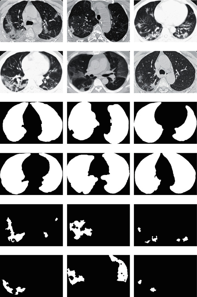
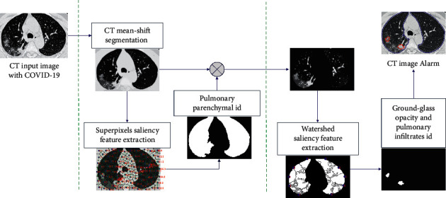
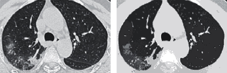
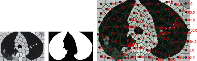
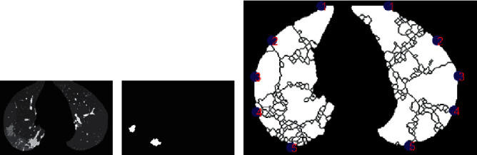
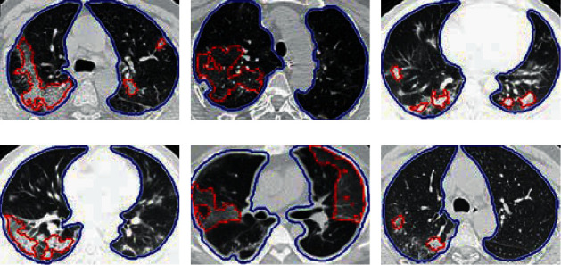
The rapid worldwide spread of the COVID-19 pandemic has infected patients around the world in a short space of time. Chest computed tomography (CT) images of patients who are infected with COVID-19 can offer early diagnosis and efficient forecast monitoring at a low cost. The diagnosis of COVID-19 on CT in an automated way can speed up many tasks and the application of medical treatments. This can help complement reverse transcription-polymerase chain reaction (RT-PCR) diagnosis. The aim of this work is to develop a system that automatically identifies ground-glass opacity (GGO) and pulmonary infiltrates (PIs) on CT images from patients with COVID-19. The purpose is to assess the disease progression during the patient's follow-up assessment and evaluation. We propose an efficient methodology that incorporates oversegmentation mean shift followed by superpixel-SLIC (simple linear iterative clustering) algorithm on CT images with COVID-19 for pulmonary parenchyma segmentation. To identify the pulmonary parenchyma, we described each superpixel cluster according to its position, grey intensity, second-order texture, and spatial-context-saliency features to classify by a tree random forest (TRF). Second, by applying the watershed segmentation to the mean-shift clusters, only pulmonary parenchyma segmentation-identified zones showed GGO and PI based on the description of each watershed cluster of its position, grey intensity, gradient entropy, second-order texture, Euclidean position to the border region of the PI zone, and global saliency features, after using TRF. Our classification results for pulmonary parenchyma identification on CT images with COVID-19 had a precision of over 92% and recall of over 92% on twofold cross validation. For GGO, the PI identification showed 96% precision and 96% recall on twofold cross validation.
Copyright © 2021 Santiago Tello-Mijares and Luisa Woo.
Conflict of interest statement
The authors declare that they have no conflicts of interest.
Figures
References
-
- World Health Organisation. 2020. Statement on the second meeting of the International Health Regulations (2005) Emergency Committee regarding the outbreak of novel coronavirus (2019-nCoV). https://www.who.int/news-room/detail/30-01-2020-statement-on-the-second-...
-
- World Health Organisation. 2020. WHO Director-General’s Opening Remarks at the Media Briefing on COVID-19 - 11 March 2020. https://www.who.int/dg/speeches/detail/who-director-general-s-opening-re....
Publication types
MeSH terms
LinkOut - more resources
Full Text Sources
Other Literature Sources
Medical
Research Materials
Miscellaneous
