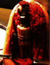
Augmented Total Elbow Arthroplasty with Femoral Strut Allograft for Revision of Prosthetic Joint Infection with Distal Humerus Bone Loss and Incomplete Union of Periprosthetic Humeral Shaft Fracture
- PMID: 33968457
- PMCID: PMC8084658
- DOI: 10.1155/2021/6661870
Augmented Total Elbow Arthroplasty with Femoral Strut Allograft for Revision of Prosthetic Joint Infection with Distal Humerus Bone Loss and Incomplete Union of Periprosthetic Humeral Shaft Fracture
Abstract
Total elbow arthroplasty (TEA) prosthetic joint infection (PJI) in the setting of distal humerus bone loss poses a challenge for restoration of function. This can be complicated by a periprosthetic humeral fracture. Revision surgery in the setting of these pathologies possesses a significant challenge, especially when two or, in this case, all three problems are treated simultaneously. We present the clinical course, operative findings, and definitive treatment with the use of an augmented total elbow arthroplasty and femoral strut allograft reinforcement in detail. A review of the literature regarding the identification and management of infected TEA with augmented prosthesis and bone allograft augmentation of humerus fractures will be outlined in this case report.
Copyright © 2021 William R. Monahan et al.
Conflict of interest statement
The authors, their immediate families, and any research foundation with which they are affiliated did not receive any financial payments or other benefits from any commercial entity related to the subject of this article.
Figures


Similar articles
-
Periprosthetic humeral fractures after total elbow arthroplasty: treatment with implant revision and strut allograft augmentation.J Bone Joint Surg Am. 2002 Sep;84(9):1642-50. J Bone Joint Surg Am. 2002. PMID: 12208923
-
Allograft-prosthetic composite reconstruction for massive bone loss including catastrophic failure in total elbow arthroplasty.J Bone Joint Surg Am. 2013 Jun 19;95(12):1117-24. doi: 10.2106/JBJS.L.00747. J Bone Joint Surg Am. 2013. PMID: 23783209
-
Proximal Humeral Replacement With Osteoarticular Allograft Prosthetic Composite in Failed Revision Total Elbow Arthroplasty With Marked Bone Loss.Tech Hand Up Extrem Surg. 2022 Jun 1;26(2):114-121. doi: 10.1097/BTH.0000000000000369. Tech Hand Up Extrem Surg. 2022. PMID: 34743164
-
[Periprosthetic humeral fractures: from osteosynthesis to prosthetic replacement].Oper Orthop Traumatol. 2019 Apr;31(2):84-97. doi: 10.1007/s00064-019-0591-y. Epub 2019 Feb 28. Oper Orthop Traumatol. 2019. PMID: 30820585 Review. German.
-
Total elbow arthroplasty for the treatment of insufficient distal humeral fractures. A retrospective clinical study and review of the literature.Injury. 2009 Jun;40(6):582-90. doi: 10.1016/j.injury.2009.01.123. Epub 2009 Apr 24. Injury. 2009. PMID: 19394013 Review.
Cited by
-
Salvage Treatment for a Periprosthetic Humeral Nonunion: A Case Report.Cureus. 2024 May 17;16(5):e60491. doi: 10.7759/cureus.60491. eCollection 2024 May. Cureus. 2024. PMID: 38883071 Free PMC article.
References
-
- Morrey M. E., Sanchez-Sotelo J., Abdel M. P., Morrey B. F. Allograft-prosthetic composite reconstruction for massive bone loss including catastrophic failure in total elbow arthroplasty. The Journal of Bone and Joint Surgery-American Volume. 2013;95(12):1117–1124. doi: 10.2106/JBJS.L.00747. - DOI - PubMed
Publication types
LinkOut - more resources
Full Text Sources

