Review
. 2021 Apr;10(4):1780-1791.
doi: 10.21037/tau-20-1441.

Narrative review of the history of microsurgery in urological practice

Affiliations
Review

Narrative review of the history of microsurgery in urological practice

Mang L Chen et al. Transl Androl Urol. 2021 Apr.

Abstract

The clinical need for magnified visualization during surgery spurred the evolution of microscope and microsuture technology. Innovative surgeons across various surgical specialties recognized the importance of utilizing and advancing these technologies. Operative microscopy allows human dexterity to perform beyond direct visual limitations. Microsurgery started in otolaryngology and ophthalmology, became popular in reconstruction and transplantation, and was then adopted in urology. Microsurgery in urology involves renal and penile revascularization, penile transplantation and free flap phalloplasty, testicular autotransplantation, reproductive tract reconstruction of the vas deferens and epididymis, varicocele repair, and sperm retrieval. By examining the peer reviewed and lay literature, this review discusses the history of microsurgery and its subsequent development as a subspecialty in urology.

Keywords: Urology; microsurgery; phalloplasty; transplantation; vasovasostomy.


Conflict of interest statement

Conflicts of Interest: All authors have completed the ICMJE uniform disclosure form (available at http://dx.doi.org/10.21037/tau-20-1441). The authors have no conflicts of interest to declare.

Figures

Figure 1
Figure 1
Photograph of Dr. Buncke’s garage laboratory (Photo courtesy of Dr. Gregory Buncke).
Figure 2
Figure 2
Photo of early microsutures developed in Dr. Buncke’s garage laboratory. The hash marks in the left panel show millimeter marks on a ruler. The left panel also shows a single needle with fine silk suture looped through the needle “eye.” The right panel shows the evolution of microsutures in which needles are molded to single-stranded microsutures (Photos courtesy of Dr. Gregory Buncke).
Figure 3
Figure 3
Amputation and reattachment of a rabbit ear. The right panel shows the first example of a successful 1 mm microvascular repair (Photos courtesy of Dr. Gregory Buncke).
Figure 4
Figure 4
Before and after photos of a patient who underwent a toe-to-thumb transplantation (Photos courtesy of Dr. Gregory Buncke).
Figure 5
Figure 5
Harry Buncke, MD and son Gregory Buncke, MD operating under the microscope (Photo courtesy of Dr. Gregory Buncke).
Figure 6
Figure 6
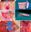
Radial forearm free flap phalloplasty: immediate postoperative photo (A) and 1-year postoperative photo (B).
Figure 7
Figure 7
Creation of a neophallus using radial forearm free flap. (A) Vaginectomy and pars fixa (PF) urethroplasty using labia minora tissue to extend the urethra from the native urethral meatus to the tip of the clitoris. (B) Dorsal nerve dissection for later coaptation with the antebrachial cutaneous nerve(s) of the radial forearm free flap. (C) Radial forearm free flap based on brachial artery, prior to pars pendulans urethroplasty and neophallus creation. Neophallus created but not yet detached from brachial artery. (D) Microsurgical nerve coaptation using 9-0 suture between the dorsal nerve of the clitoris and the antebrachial cutaneous nerve (left panel) and microvascular anastomosis of cephalic vein to greater saphenous vein (right panel).
Figure 8
Figure 8
Microphotographs of the Berger triangulation invagination epididymovasostomy technique for vasectomy reversal (30× magnification). Left panel: Three 10.0 microsutures placed in a single epididymal tube as “vest” sutures. The epididymal tubule is then punctured and the microneedles advanced and placed inside-out into the vas deferens (right panel). Once the sutures are tied, the epididymal tubule is “invaginated” or advanced into the vas deferens lumen.
Figure 9
Figure 9
Subinguinal spermatic cord dissection and pampiniform vein ligation during varicocelectomy (10× magnification). Left panel: exposure of subinguinal spermatic cord with gonadal veins; Right panel: spermatic cord after ligation of gonadal veins. Vas deferens (labelled in yellow) and its veins and artery are excluded from the ligation.

References

    1. Nylen COS. Något om de senaste 25 årens utveckling inom klinisk vestibularforskning, särskilt rörande labyrintfistelsymtom och lägenystagmus. Uppsala, Leipzig, A.-b: Lundequistska bokhandeln; 1942.
    1. Dohlman GF. Carl Olof Nylen and the birth of the otomicroscope and microsurgery. Arch Otolaryngol 1969;90:813-7. 10.1001/archotol.1969.00770030815025 - DOI - PubMed
    1. Schultheiss D, Denil J. History of the microscope and development of microsurgery: a revolution for reproductive tract surgery. Andrologia 2002;34:234-41. 10.1046/j.1439-0272.2002.00499.x - DOI - PubMed
    1. Berland T, Perritt RA. Living with your eye operation. New York: St. Martin's Press, 1974.
    1. Swolin K. Contribution to the surgical treatment of female sterility. Experimental and clinical studies. Acta Obstet Gynecol Scand 1967;46 Suppl:14-20. - PubMed