

The current value of quantitative shear wave sonoelastography in parotid gland tumors
- PMID: 33968689
- PMCID: PMC8102225
- DOI: 10.21037/gs-20-837
The current value of quantitative shear wave sonoelastography in parotid gland tumors
Abstract
Background: The preoperative differentiation between salivary gland tumor entities using computed tomography, magnetic resonance imaging (MRI) and ultrasound (US) is still limited. Biopsies are often regarded as indispensable for properly characterizing these various lesions. The aim of this study was to analyze the value of acoustic radiation force impulse (ARFI) sonoelastography as an US differentiation tool when examining parotid gland (PG) lesions.
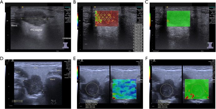
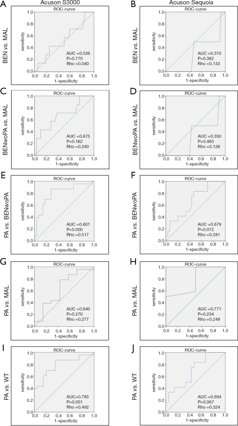
Methods: We included 104 patients with PG masses in this study, employing two different US devices using quantitative ARFI-sonoelastography (Siemens Acuson-S3000, n=59; Siemens Acuson-Sequoia, n=45). The ability of sonoelastographic measurements to differentiate between different neoplasms was compared and analyzed for both US machines.
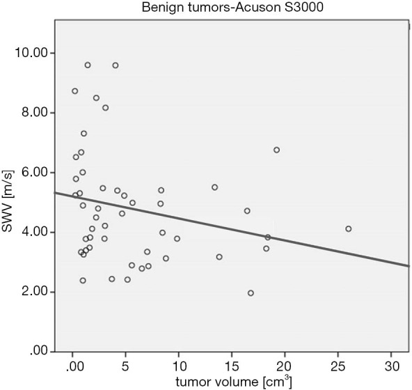
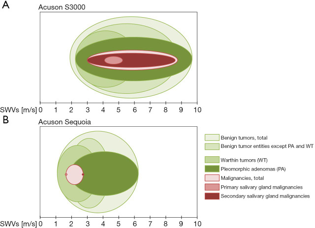
Results: Quantitative shear wave sonoelastography is limited in its ability to reliably differentiate between tumor entities of the PG as a stand-alone parameter. Measurement results were unsystematically distributed and not transferable between the two US devices. A significant differentiation of benign and malignant lesions was not possible with either US machine (S3000: P=0.770, Sequoia: P=0.382). A differentiation between pleomorphic adenomas (PA) and Warthin tumors was only possible with the Acuson S3000 system (P=0.001, Spearman-Rho =0.492, sensitivity 73.9%, specificity 65.0%).
Conclusions: A reliable identification and differentiation of PG tumors as well as clinical treatment decisions cannot be made with the sole use of ARFI-sonoelastography. The results emphasize the device-dependence and high error-proneness of this US technique when examining lesions of the PG.
Keywords: Ultrasound (US); acoustic radiation force impulse (ARFI); parotid gland (PG); salivary gland tumors; sonoelastography.
2021 Gland Surgery. All rights reserved.
Conflict of interest statement
Conflicts of Interest: All authors have completed the ICMJE uniform disclosure form (available at http://dx.doi.org/10.21037/gs-20-837). The authors have no conflicts of interest to declare.
Figures
Similar articles
-
Evaluation of parotid gland lesions with standard ultrasound, color duplex sonography, sonoelastography, and acoustic radiation force impulse imaging - a pilot study.Ultraschall Med. 2012 Jun;33(3):283-8. doi: 10.1055/s-0031-1299130. Epub 2012 Apr 13. Ultraschall Med. 2012. PMID: 22504938
-
Initial Experience With Ultrasound Elastography for Diagnosis of Major Salivary Gland Lesions.J Ultrasound Med. 2016 Dec;35(12):2597-2606. doi: 10.7863/ultra.15.11093. Epub 2016 Oct 25. J Ultrasound Med. 2016. PMID: 27872416
-
Clinical Utility of Qualitative Elastography Using Acoustic Radiation Force Impulse for Differentiating Benign from Malignant Salivary Gland Tumors.Ultrasound Med Biol. 2021 Feb;47(2):279-287. doi: 10.1016/j.ultrasmedbio.2020.10.007. Epub 2020 Nov 12. Ultrasound Med Biol. 2021. PMID: 33189412
-
Sonoelastography for differential diagnosis between malignant and benign parotid lesions: a meta-analysis.Eur Radiol. 2019 Feb;29(2):725-735. doi: 10.1007/s00330-018-5609-6. Epub 2018 Jul 10. Eur Radiol. 2019. PMID: 29992386 Free PMC article. Review.
-
Sonoelastography techniques in the evaluation and diagnosis of parotid neoplasms.Eur Radiol. 2012 May;22(5):966-9. doi: 10.1007/s00330-012-2401-x. Epub 2012 Feb 26. Eur Radiol. 2012. PMID: 22367472 Review.
Cited by
-
Assessment of Parotid Gland Tumors by Means of Quantitative Multiparametric Ultrasound (mpUS).Diagnostics (Basel). 2022 Dec 21;13(1):12. doi: 10.3390/diagnostics13010012. Diagnostics (Basel). 2022. PMID: 36611304 Free PMC article.
-
Salivary gland pathologies: evolution in classification and association with unique genetic alterations.Eur Arch Otorhinolaryngol. 2023 Nov;280(11):4739-4750. doi: 10.1007/s00405-023-08110-w. Epub 2023 Jul 13. Eur Arch Otorhinolaryngol. 2023. PMID: 37439929 Free PMC article. Review.
References
LinkOut - more resources
Full Text Sources